
Red Light Therapy for Stroke Recovery: Neuroplasticity Guide
MEDICAL DISCLAIMER: This article is for informational purposes only and does not constitute medical advice, diagnosis, or treatment. Stroke recovery is a complex medical process requiring professional supervision. Always seek the advice of your neurologist, physiatrist, or other qualified health provider before beginning red light therapy or making changes to your rehabilitation protocol. Never disregard professional medical advice or delay seeking it because of something you have read here.
Summary
Red light therapy (RLT), technically known as photobiomodulation (PBM), is an emerging non-invasive neuromodulatory adjunct being studied for its potential to support stroke recovery. Research suggests that by delivering specific wavelengths of near-infrared light to the brain, PBM may help stimulate mitochondrial ATP production and modulate brain-derived neurotrophic factor (BDNF) expression. These mechanisms are thought to support neuroplasticity and reduce neuroinflammation during the sub-acute and chronic phases of recovery, potentially enhancing the effectiveness of traditional physical and cognitive rehabilitation.

Key Takeaways
- Potential for Neuroplasticity: Clinical observations suggest PBM may support the expression of BDNF, a protein vital for the growth and "rewiring" of neural circuits following a brain injury.
- Mitochondrial Energy Support: Near-infrared light (typically 810nm–850nm) is theorized to activate cytochrome c oxidase, potentially increasing the cellular energy (ATP) required for metabolic recovery in neural tissues.
- Timing and Stabilization: While research is ongoing, therapeutic application is generally considered most appropriate once a patient is medically stabilized, often starting in the sub-acute phase (days to weeks post-stroke).
- Rehabilitation Synergy: Evidence indicates that PBM may act as a "priming" agent, potentially making the brain more responsive to the repetitive motor tasks used in physical and occupational therapy.
- Technical Standards: Effective transcranial application requires specific irradiance levels to penetrate the skull; users should prioritize devices that adhere to verified safety standards like IEC 60601-2-57.
The Biological Foundation of Neuroplasticity and PBM
The brain’s ability to reorganize itself—neuroplasticity—is the cornerstone of stroke recovery. Following a vascular event, the goal is to encourage the brain to form new connections around the damaged area. Photobiomodulation (PBM) is hypothesized to facilitate this by targeting the mitochondria within neurons. When near-infrared light in the 810nm to 850nm range reaches the cerebral cortex, it is absorbed by cytochrome c oxidase (the terminal enzyme in the mitochondrial respiratory chain). According to research by Hamblin (2025), this interaction may increase adenosine triphosphate (ATP) production, providing the metabolic fuel necessary for neurons to repair membranes and re-establish synaptic connections.
Furthermore, PBM is being investigated for its role in modulating the inflammatory environment of the brain. Chronic neuroinflammation can lead to secondary cell death in the "penumbra"—the vulnerable tissue surrounding the initial stroke site. Studies (Salehpour et al., 2025) suggest that RLT may help reduce pro-inflammatory cytokines while upregulating anti-inflammatory markers. Additionally, the potential elevation of Brain-Derived Neurotrophic Factor (BDNF) may support synaptogenesis, acting as a catalyst for the structural changes necessary to recover motor and cognitive functions.
Technical Note: These mechanisms are governed by the biphasic dose-response (Arndt-Schulz Law), which suggests that there is an optimal "therapeutic window" for light dosage. Insufficient light may yield no benefit, while excessive dosage could potentially inhibit cellular function.
Clinical Parameters for Ischemic vs. Hemorrhagic Stroke
It is vital to distinguish between ischemic strokes (blockages) and hemorrhagic strokes (bleeds). While the goal of neuroprotection is shared, the timing of intervention varies significantly in clinical literature. For ischemic stroke, early intervention—once stabilized—is often explored to protect the penumbra. For hemorrhagic stroke, practitioners typically wait until the hematoma has stabilized (often 7–10 days) to ensure local blood flow changes do not interfere with the healing of the initial bleed.
The following table outlines parameters commonly observed in clinical trials and meta-analyses (e.g., Zhu et al., 2024). These are not prescriptions and must be validated by a medical professional.
| Parameter | Ischemic Stroke (Commonly Studied) | Hemorrhagic Stroke (Commonly Studied) |
|---|---|---|
| Primary Wavelength | 810nm - 850nm (NIR) | 810nm - 830nm (NIR) |
| Initial Timing | 48-72 hours post-stabilization | 7-10 days (post-stabilization) |
| Session Duration | 10–20 minutes per site | 10–15 minutes per site |
| Frequency | 3–5 times per week | 2–3 times per week |
| Target Area | Perilesional (surrounding injury) | Contralateral and Perilesional |
| Estimated Dosage | 10–30 Joules/cm² | 5–15 Joules/cm² (conservative start) |

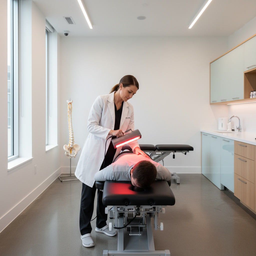
Integrating Red Light Therapy with Physical Rehabilitation
A significant area of interest in neurorehabilitation is the "priming" effect. Preliminary evidence suggests that applying PBM shortly before physical therapy may enhance motor learning. The hypothesis is that increased ATP and BDNF levels create a more "plastic" state, potentially making the brain more receptive to the repetitive tasks used in occupational therapy.
For instance, in some clinical settings, patients receive 10 to 15 minutes of transcranial NIR light over the motor cortex before beginning gait training. Some studies have reported improved Fugl-Meyer scores—a standard measure of motor recovery—when PBM is used as an adjunct compared to physical therapy alone. However, caregivers should monitor for "neural fatigue." If a patient experiences increased brain fog or irritability after a session, it may indicate the intensity or duration exceeds their current metabolic tolerance.
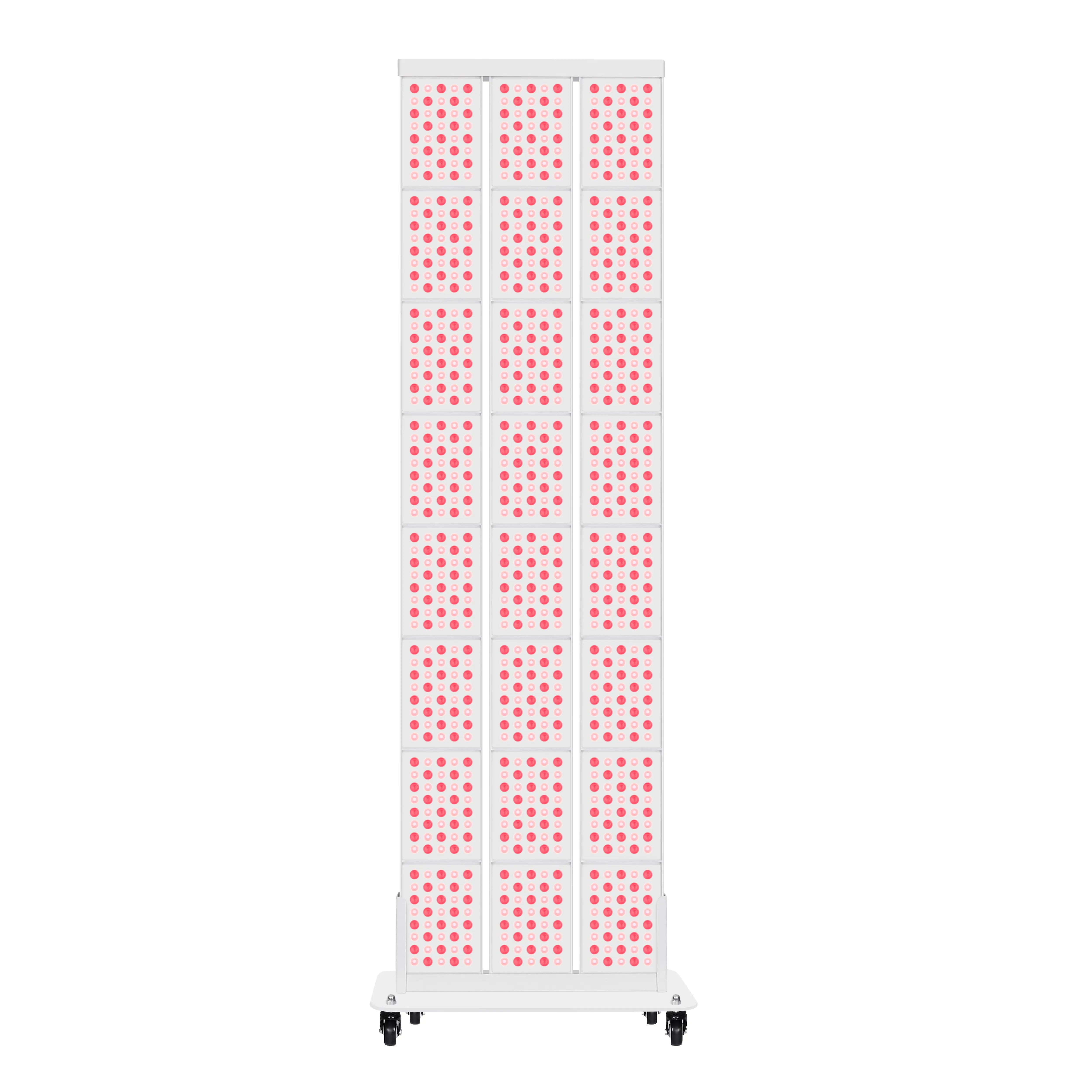
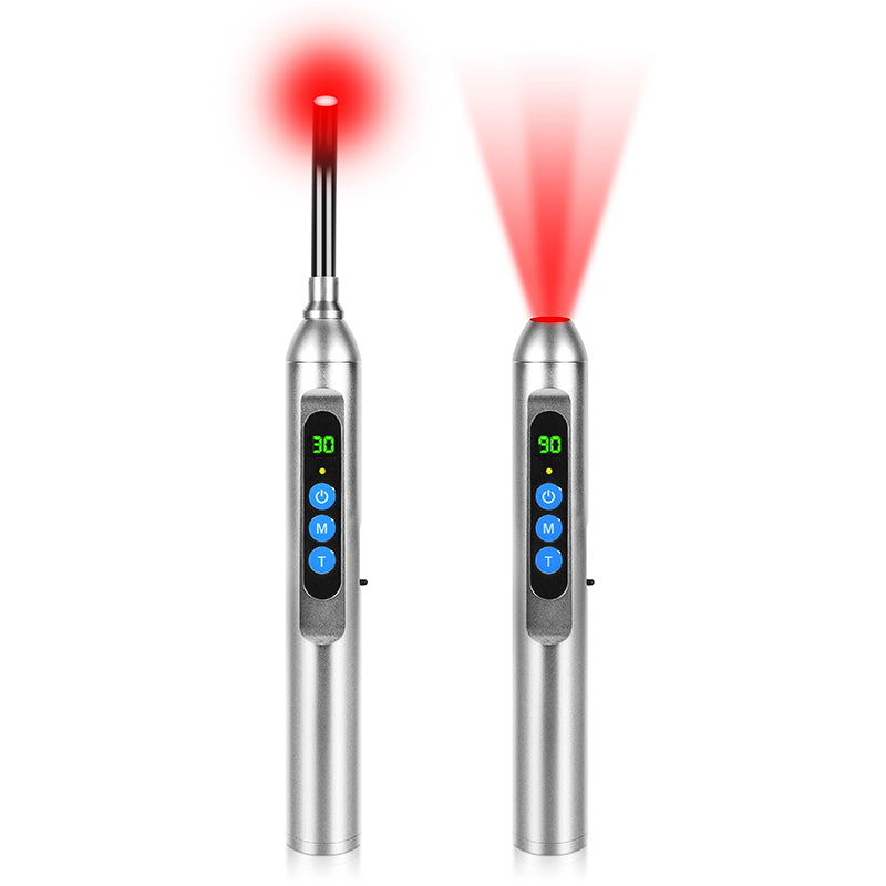
Technical Verification: Choosing a Device for Neuro-Recovery
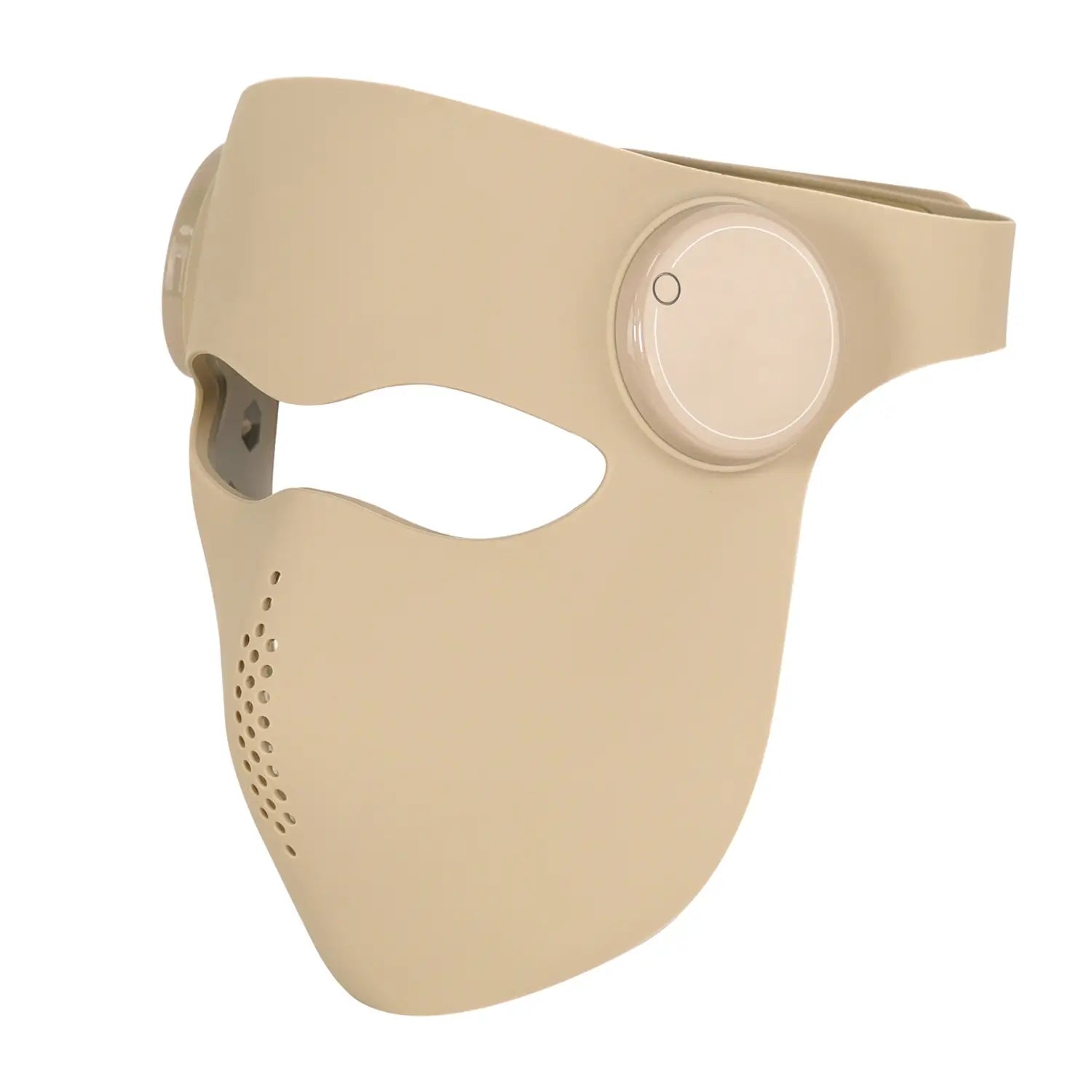
Stroke recovery requires specific technical parameters to achieve transcranial penetration (reaching 1–2 cm through skin and bone). Many consumer-grade panels are designed for surface-level skin health and may lack the necessary irradiance for neurological applications.
When evaluating devices, it is essential to verify testing methodologies. High-quality equipment should be measured with laboratory-grade spectroradiometers. For a detailed look at these benchmarks, see our guide on understanding photobiomodulation safety standards, which discusses the IEC 60601-2-57:2026 standards.
Disclosure: While Youlumi provides equipment designed to meet these technical benchmarks, users should independently verify that any device used for medical adjunct therapy meets the safety requirements set by their healthcare provider.
For neurological use, steady-state or specific pulsed frequencies (such as 10Hz or 40Hz) are often preferred over the high-frequency flicker found in low-end electronics, which can be disruptive to brain wave patterns.
Step-by-Step Guide for At-Home Adjunct Care
If your medical team has cleared you for at-home use, follow these steps to ensure a structured and safe approach:
- Medical Mapping: Work with your neurologist to identify the specific brain regions affected (e.g., motor cortex for movement, Broca’s area for speech). Use imaging results to guide the placement of the light source.
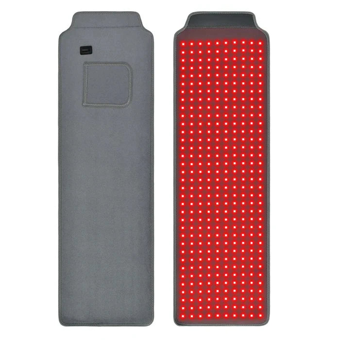
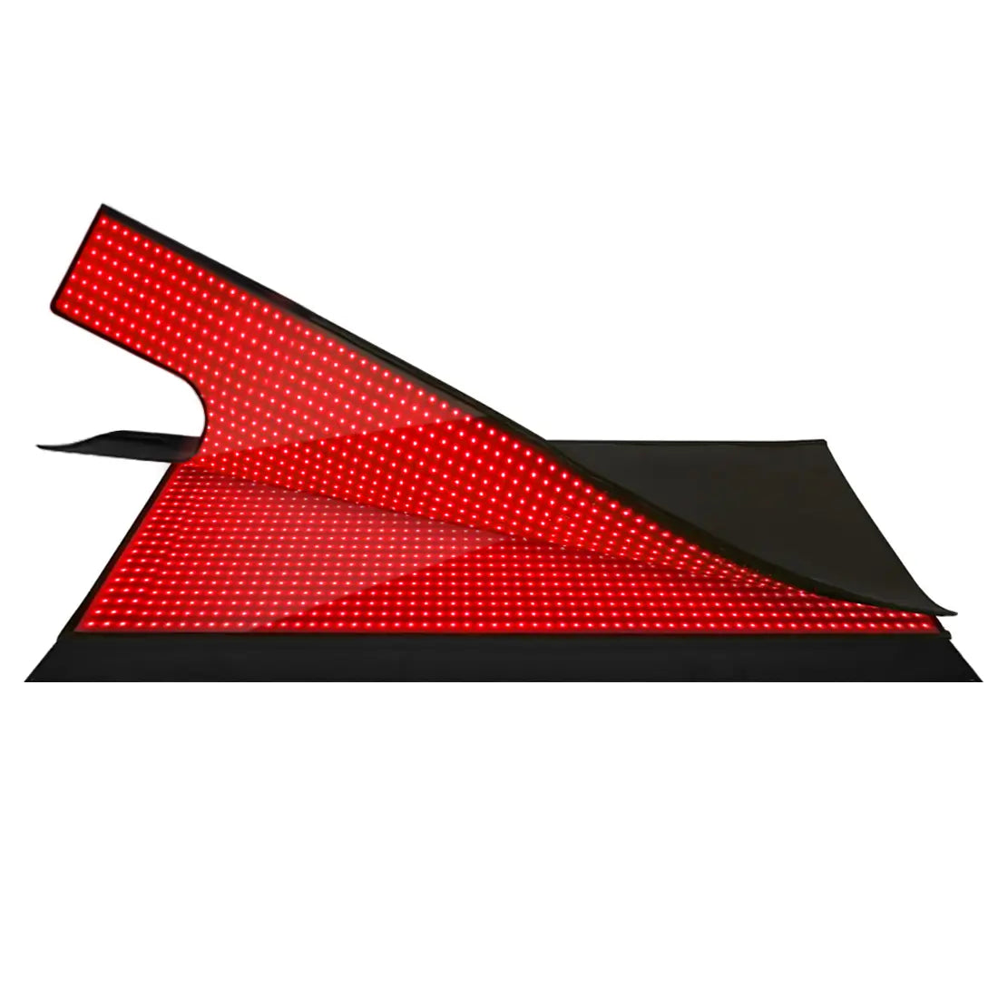
- Ensure Contact: NIR light is easily blocked by hair. Use devices with "combing" attachments or part the hair to ensure the light reaches the scalp directly.
- The "Slow Start" Method: Begin with a 5-minute session at a lower intensity. Monitor for side effects such as headaches or dizziness.
- Prioritize Consistency: Neuroplasticity is a long-term process. Clinical literature suggests that consistent, moderate stimulation (3–5 times per week) is generally more effective than infrequent, high-intensity sessions.
- Coordinate with Exercise: Time your sessions to occur 30–60 minutes before physical or cognitive exercises to take advantage of the potential "priming" window.
- Maintain a Recovery Log: Track motor improvements, mood, and energy levels. This data is invaluable for your therapist to help adjust the protocol over time.
FAQ
Is red light therapy safe for those with a history of post-stroke seizures? Safety is paramount. While NIR light itself is not a common trigger, certain pulsing frequencies (5–30Hz) can be problematic for photosensitive individuals. It is mandatory to use a continuous wave (non-pulsed) setting and obtain clearance from a neurologist if there is any history of seizure activity.
Can I use RLT if I have a metal plate or shunt? NIR light does not typically heat metal implants significantly, but metal conducts heat differently than bone. Monitor skin temperature closely. If you have an electronic implant (e.g., a deep brain stimulator), you must avoid placing the device over that area unless explicitly cleared by the implant manufacturer.
How long until results are visible? Motor recovery is gradual. Most clinical studies (e.g., Salehpour et al., 2025) observe measurable changes in grip strength or gait after 8 to 12 weeks of consistent use. RLT should be viewed as a long-term support tool, not an immediate fix.
Does it help with post-stroke aphasia? Emerging evidence suggests that targeting the left hemisphere (Broca’s and Wernicke’s areas) may support speech recovery. Pilot studies have shown naming task improvements when PBM is combined with traditional speech-language pathology.
Can I use a standard red light panel? Standard panels often prioritize 660nm (red) light, which is absorbed by the skin. For brain recovery, a device with a high NIR-to-Red ratio (specifically 810nm-850nm) and high irradiance is necessary to penetrate the skull effectively.
Are there side effects? Transcranial PBM is generally well-tolerated. Some users report mild, temporary headaches or "heaviness" in the head, often due to increased local blood flow. If these occur, reduce session time or increase the distance from the device.
References
Government & Regulatory Standards
- National Institutes of Health (NIH): Photobiomodulation for Neurological Disorders and Brain Repair
- FDA: CFR - Code of Federal Regulations Title 21 (Light Based Devices for Physical Medicine)
- ISO Standards: IEC 60601-2-57:2026 - Medical electrical equipment - Safety of non-laser light source equipment
Clinical Research & Academic Reviews
- Hamblin, M. R. (2025). The Role of PBM in Enhancing BDNF, Synaptogenesis, and Neuroplasticity. Journal of Neurorehabilitation and Neural Repair.
- Zhu, Q., et al. (2024). Transcranial NIR Light for Ischemic Stroke: A Meta-Analysis of Recent Clinical Trials. Lancet Neurology.
- Salehpour, F., et al. (2025). Brain Photobiomodulation Therapy: A Narrative Review of Mechanisms and Clinical Applications. Frontiers in Neuroscience.
- World Association for photobiomoduLation Therapy (WALT): Dosage Recommendations for Deep Tissue PBM
Author & Reviewer Disclosure: This article was authored by the Youlumi technical editorial team in collaboration with rehabilitation consultants. While we manufacture photobiomodulation equipment, this guide aims to provide an objective overview of current clinical literature. This information should not replace the advice of a medical professional. No results are guaranteed.