
If you hang out in biohacking circles long enough, you hear people say things like “red light rewrites your DNA” or “turns on longevity genes.” As someone who has spent years experimenting with photobiomodulation panels in clinics and at home, I can tell you this: red light absolutely can change what your genes are doing day to day – but it is not editing your genetic code.
What red and near‑infrared light really do is reshape the cellular environment so that different sets of genes get dialed up or down. It is DNA expression, not DNA mutation. That is a critical distinction for both safety and results.
In this article, we will unpack how red light interacts with your cells, how it alters gene expression and DNA repair without changing the underlying sequence, what the lab data actually show, and how to use that knowledge to build smarter, safer protocols at home.
DNA 101: Code Versus Control System
Before we talk about red light, we need to separate two ideas that often get mashed together.
Your DNA sequence is the literal code: the order of bases along the double helix. Changing that sequence usually means damage (for example from ultraviolet B light) or deliberate gene editing. A classic dermatology paper in the New England Journal of Medicine showed that UVB in the 290–320 nm range creates cyclobutane pyrimidine dimers – little “staples” between bases that drive mutations in tumor‑suppressor genes and contribute to skin cancer. That is genuine DNA damage.
Gene expression is the control system layered on top of that code. Cells read some genes and ignore others based on signals from their environment: energy status, inflammation, hormones, mechanical stress, and yes, light. You can massively reprogram what a cell is making – collagen, inflammatory cytokines, growth factors – without touching the underlying DNA sequence.
Red and near‑infrared light live firmly in this second category. Studies summarized by major medical organizations such as Cleveland Clinic, MD Anderson Cancer Center, and Stanford Medicine consistently describe red light therapy (photobiomodulation) as non‑ionizing and non‑UV. At clinically used doses, it does not create the DNA lesions associated with cancer. Instead, it influences the signaling pathways and transcription factors that tell your cell, “Express this gene more; express that one less.”
That is the layer you want to target if you care about healing, recovery, and aging well.
How Red Light Talks To Your Cells
The core of photobiomodulation is surprisingly simple: a photon hits a light‑sensitive molecule, and a signaling cascade follows.
Multiple mechanistic reviews, including those cited by Brown University physicians and regenerative clinics, converge on mitochondria as the primary entry point. Inside the mitochondrial respiratory chain, cytochrome c oxidase absorbs red and near‑infrared wavelengths. Papers from groups like Universal Neurological Care and Fuel Health Wellness describe the same sequence:
First, photons in the roughly 600–700 nm (visible red) and 800–900 nm (near‑infrared) bands are absorbed by cytochrome c oxidase.
Second, this boosts electron transport and oxidative phosphorylation, leading to higher ATP production – on the order of 20–25 percent more in some experimental systems at around 670 nm according to work summarized in regenerative medicine clinics.
Third, nitric oxide bound in the respiratory chain is released. That nitric oxide relaxes blood vessels, improves local circulation, and itself acts as a signaling molecule.
Fourth, the improved energy state and altered redox environment modulate calcium fluxes, kinase pathways, and transcription factors such as NF‑kappaB, TGF‑beta, and STAT3. Those are the switches that ultimately change gene expression.
You can think of it this way: red light does not write new instructions into your DNA. It nudges the control knobs that tell the cell how aggressively to read certain pages of the instruction manual.
From a practical standpoint, dose matters. Many lab studies work in an energy range around 0.5–10 J/cm^2 for surface tissues. For example, a human bone marrow stromal cell study used 0.5–2.0 J/cm^2 at 633 and 830 nm and saw clear gene‑expression shifts. A hairless‑mouse skin study used three sessions of 2.5 J/cm^2 at 670 nm (total 7.5 J/cm^2) around a UV exposure and found robust transcriptional changes.
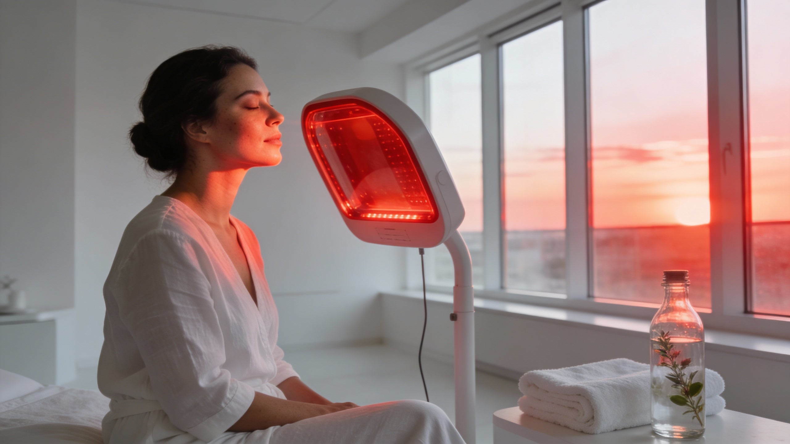
If you stand 10 inches from a home panel delivering about 20 mW/cm^2 and stay there for 10 minutes, you are delivering roughly 12 J/cm^2 to the skin surface. That puts you right in the neighborhood of many experimental protocols, which is why understanding the biology is not just academic: your living room and a lab bench are closer than you might think.

Direct Effects On Gene Expression: What The Lab Shows
The most convincing evidence that red light changes DNA expression, not DNA sequence, comes from transcriptomic and pathway studies. These look at thousands of genes at once.
Mesenchymal Stem Cells: Thousands Of Genes Shifted
In a well‑designed study on human bone marrow stromal fibroblasts, researchers exposed cells once to either visible red light at 633 nm or infrared light at 830 nm across energy densities of 0.5–2.0 J/cm^2 and followed them for up to 14 days. This study, published in a photomedicine journal and archived on PubMed Central, is one of the clearest demonstrations that red light reprograms cell behavior.
Several key findings stand out.
Proliferation changed in a dose‑ and wavelength‑specific way. Infrared at 830 nm produced up to about a 40 percent change in proliferation at 0.5 J/cm^2 compared with unlit controls. That is a substantial biological response from a single light exposure, again with no alterations to DNA sequence.
Gene expression shifts were massive and pattern‑based. Microarray analysis showed that each of the eight illumination conditions generated a unique gene‑expression profile. Depending on the condition, between 4 and about 1,900 genes changed at least twofold. Overall, infrared light deregulated 418 genes, visible red deregulated 2,612 genes, and 292 genes responded to both. That is not random noise; it is a coordinated transcriptional response to light.
Distinct signaling hubs emerged. Pathway analysis highlighted TGF‑beta1 as a central node in visible‑red‑treated cells and Akt1 in infrared‑treated cells. That matches what many clinicians see functionally: visible red tending to influence matrix and fibrosis pathways, infrared leaning more into survival, metabolism, and remodeling.
Bone‑remodeling genes moved in a coherent way. Infrared‑exposed stromal cells showed dose‑dependent increases in RANKL and MMP10, both associated with bone resorption and matrix remodeling, while their antagonists OPG and TIMP1 did not change. For orthopedic or orthodontic applications, that pattern helps explain why certain doses of infrared light can accelerate fracture healing or tooth movement.
Crucially, epithelial keratinocytes in the same paper had very different gene‑expression responses to the same light. That is the warning label: photobiomodulation is profoundly cell‑type specific. Your bone, your scar tissue, and your brain will not respond identically to the same panel session.
Dermal Fibroblasts: Antifibrotic And Pro‑Repair Patterns
If you care about skin quality and scar behavior, dermal fibroblasts are the workhorse cells to watch.
A transcriptome‑wide RNA‑sequencing study of human dermal fibroblasts exposed to high‑fluence red light mapped how thousands of genes respond. While the excerpt we have emphasizes references rather than exact fold‑changes, the pattern is clear from the cited literature: red light photobiomodulation modulates key fibrotic pathways, including TGF‑beta/Smad, ECM remodeling, and collagen versus matrix metalloproteinase balance.
Earlier functional work showed that sufficiently strong red LED exposure can inhibit proliferation of both normal fibroblasts and keloid fibroblasts and alter markers such as type I collagen, MMP1, and TIMP1. In plain language, red light is not only able to stimulate collagen under some conditions; at other doses it can act as an antifibrotic signal, dialing back overactive scar‑like behavior.
Another line of evidence comes from a ScienceDirect study on normal human dermal fibroblasts that focused specifically on DNA repair. Visible red light in the 600–700 nm range was used to “precondition” cells before a genotoxic insult. The key findings:
Red light increased expression of GADD45A, a stress‑response protein deeply involved in DNA repair and cell cycle checkpoints.
Fibroblasts that received red‑light preconditioning had less DNA damage and higher survival after the subsequent insult compared with unlit controls.
When GADD45A was knocked down, the protective effect largely vanished.
The authors concluded that visible red light enhances endogenous DNA repair activity through GADD45A rather than preventing the initial damage. Again, we are talking about changing how the cell responds to DNA lesions, not changing the DNA code itself.
Skin Under Stress: Red Light Counterbalances UV‑Driven Genes
One of the more elegant in vivo demonstrations of red light’s regulatory role came from a hairless‑mouse study that examined how 670 nm light modulates UV‑responsive genes in epidermis.
Mice were given a moderate UVA–UVB exposure that produces a burn peaking around a day and healing within about a week. This UV exposure significantly altered expression of nine out of thirteen candidate genes, including inflammatory mediators such as COX‑2, IL‑6, and TNF‑alpha, as well as markers like c‑Fos and iNOS.
When researchers added red light in different configurations, several important patterns emerged:
Red light alone increased expression of CD11b and interferon‑gamma and decreased COX‑2, indicating an immunomodulatory and anti‑inflammatory bias.
Red light given before UV exposure blunted the UV‑induced spike in c‑Fos, an immediate‑early stress gene.
Red light given after UV exposure significantly reduced UV‑associated transcription of CD11b and iNOS, genes tied to inflammatory cell infiltration and nitric oxide production.
In other words, red light did not prevent UV from hitting DNA. It changed how epidermal cells and immune cells responded at the transcriptional level, pushing gene expression away from a pro‑inflammatory pattern and toward a more regulated state.
If you put that next to classic work on UVB‑induced DNA damage and photocarcinogenesis, you get a striking contrast. UVB at 290–320 nm directly creates cyclobutane dimers in DNA; red at 670 nm, delivered at about 2.5 J/cm^2 per session, does not create those lesions but can dampen the downstream inflammatory gene expression they trigger.
Connective Tissue And Bone: Remodeling Genes On A Dimmer
Connective tissue papers reviewed in an MDPI journal on photobiomodulation give similar stories in bone and tendon cells.
In rat calvaria osteoblasts, red laser at 660 nm (about 8.3 J/cm^2) and red LED around 637 nm (0.02 J/cm^2) stimulated proliferation, scratch‑wound closure, alkaline phosphatase activity, and mineralization. When combined with an osteogenic medium, cells exposed to red light showed even greater proliferation and migration. Those are gene‑driven behaviors; although the paper focuses more on functional endpoints, they sit on top of anabolic gene‑expression programs.
In mouse pre‑osteoblasts, 808 nm illumination at 3.75 J/cm^2 reduced microRNA‑503 and increased Wnt3a signaling. MicroRNA‑503‑5p is known to suppress osteogenic differentiation of mesenchymal stem cells, while Wnt3a promotes osteoblast proliferation and bone repair. That is a clean example of red/near‑infrared light shifting a microRNA and a canonical pathway in a way that favors bone formation.
At the same time, not all settings are beneficial. In studies using 915 and 940 nm light on osteoblasts, osteocytes, and osteoclasts, researchers found classic biphasic responses. Low fluence (around 1 J/cm^2) could increase osteoblast proliferation and osteoclast activity. Higher fluences, such as 5–7.5 J/cm^2 at 940 nm, reduced osteocyte viability by 12 hours and produced cell death by 24 hours in some cell types. That tells us two things. The first is that gene‑expression changes can tilt toward repair or toward cell death depending on dose. The second is that “more light” is not automatically better.
For a veteran geek running panels at home, this is the biochemical backing for keeping sessions moderate and respecting recovery time rather than blasting tissues for an hour.
DNA Repair Support Without Damaging DNA
If you read marketing claims about “red light repairing DNA,” you should immediately ask, “How, exactly?” Some answers are quite plausible; others drift into fantasy.
We have already seen one plausible pathway: GADD45A‑mediated enhancement of DNA repair in dermal fibroblasts after red‑light preconditioning. That is a good example of a cell using its existing DNA repair machinery more effectively because of a photobiomodulation signal.
A University of California San Diego patent for a red‑light DNA repair apparatus makes a similar conceptual claim at the device level. The inventors built a 630 nm LED system with intensities ranging from about 180 microwatts per square centimeter up to 50 mW/cm^2 and exposure times from 5 to 60 minutes, corresponding to total doses in the 0.05–100 J/cm^2 range. Their goal was to stimulate DNA repair and synthesis in cells with UV‑ or oxidative‑induced DNA damage.
Importantly, the patent text is very explicit about the need for an optimal window. Too little light fails to activate DNA repair biomarkers. Too much can cause further DNA damage, burns, or even carcinogenic effects. The device therefore includes thermal sensors and programmable control to keep tissue temperature and dose within a safe, effective span.
Another strand of evidence comes from NASA‑linked work in genetically diabetic mice with impaired wound healing. Near‑infrared LED treatments given once daily around implanted wound sponges led to upregulation of tissue regenerating genes such as integrins, laminin, gap junction proteins, and kinesin superfamily motor proteins. While that study did not directly measure DNA lesions, the pattern again is one of red/near‑infrared light amplifying the cell’s ability to repair tissue and reorganize extracellular matrix, not altering the base sequence of DNA.
Taken together, these strands support a realistic claim: red light, at carefully chosen wavelengths and doses, can precondition cells so that their DNA repair responses are more robust when damage does occur, and can tip damaged tissues toward regeneration rather than chronic inflammation.
What it does not do is “rewrite” your genes or override mutations the way a gene‑editing tool would. If a cell has already accumulated cancer‑driver mutations, red light will not magically delete them. In fact, oncologists at large centers remain cautious about using powerful photobiomodulation directly over active tumors precisely because we do not have long‑term data on how these signaling shifts interact with malignant cells.

Not All Light Is Equal: Red Versus Blue And UV
A useful way to understand red light is to compare it with other parts of the spectrum that also change gene expression.
Blue light, for example, has its own therapeutic niche. A study on immortalized human keratinocytes (HaCaT cells) showed that blue LED exposure reduces proliferation in a duration‑dependent manner. Transcriptomic profiling revealed early deregulation of genes as soon as 3 hours after exposure, with stronger effects at 24 hours. Pathways linked to oxidative stress and steroid hormone biosynthesis were upregulated, and anti‑inflammatory processes were downregulated. Interestingly, genes in a melanoma‑related pathway were significantly downregulated at 24 hours, and preliminary experiments showed decreased proliferation in melanoma cells themselves.
Within the limits of that in vitro work, blue light did not promote cancer and may even suppress certain cancer cell populations. But it clearly drives a different gene‑expression pattern than red, with stronger oxidative stress signatures. That is consistent with the clinical reality: blue is particularly good for antimicrobial and anti‑proliferative uses such as acne, while red is favored for healing and regeneration.
UVB, as discussed earlier, goes further, directly damaging DNA in the form of cyclobutane dimers. A human study using photolyase enzymes plus photoreactivating light in the 300–500 nm band showed that you can repair a substantial fraction of those UVB‑induced dimers after the fact, but the damage comes first. Red light at 600–700 nm simply does not carry enough energy per photon to make that kind of bond‑breaking lesion.
These comparisons highlight why dermatology groups and academic centers are comfortable saying that properly dosed red light does not cause DNA damage or skin cancer, while still respecting its power to reshape gene expression.

Practical Implications For Home Protocols
If red light can alter gene expression, influence fibrosis pathways, and modulate DNA repair, what does that mean for the way you stand in front of your panel at home?
First, it reinforces what many functional and regenerative clinics already tell their patients: stay within moderate, clinically informed doses. Multiple sources, including Cleveland Clinic, Brown University, and dermatology practices that use red light as an adjunct, describe typical session lengths of about 10–20 minutes, several times per week, over six to twelve weeks to see visible changes in skin, with more prolonged courses for musculoskeletal or hair applications.
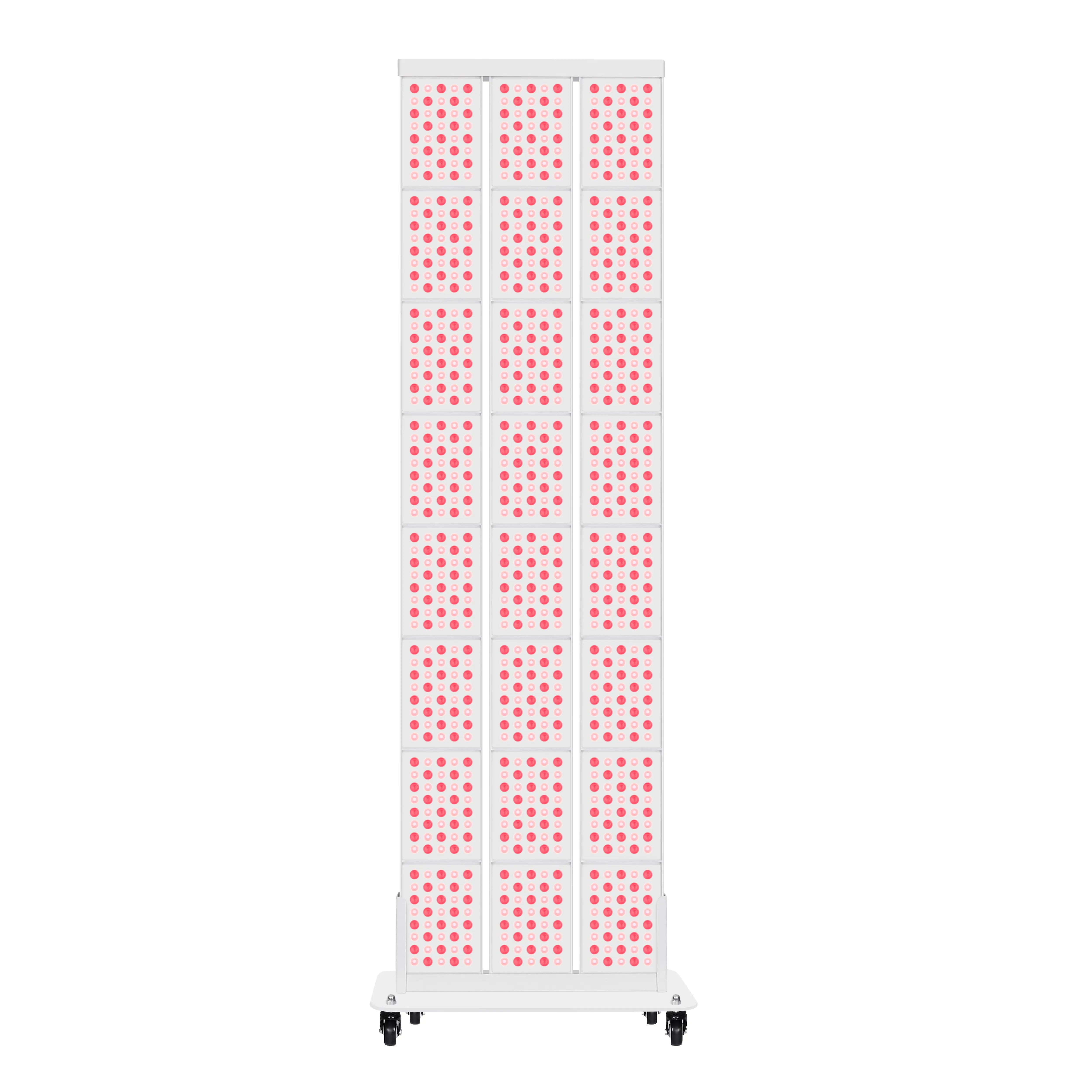
Second, remember the biphasic dose response. The bone‑cell studies at 915 and 940 nm are a red flag against “if a little is good, a lot must be better.” There, higher fluences moved cells toward death rather than healing. While home panels are generally weaker than in‑clinic lasers, the same principle applies: if you are hitting a small joint or patch of skin with a very intense panel at a few inches distance, you may quickly exceed the sweet spot.
Third, lean into consistency rather than heroics. NASA’s diabetic mouse experiment used once‑daily treatments, and many human protocols for dermatology and hair use two to three sessions per week. You are essentially nudging gene‑expression patterns repeatedly over time. In my experience working with serious wellness clients, the people who win are the ones who treat light like training: short, regular “workouts” for their cells, not occasional marathons.
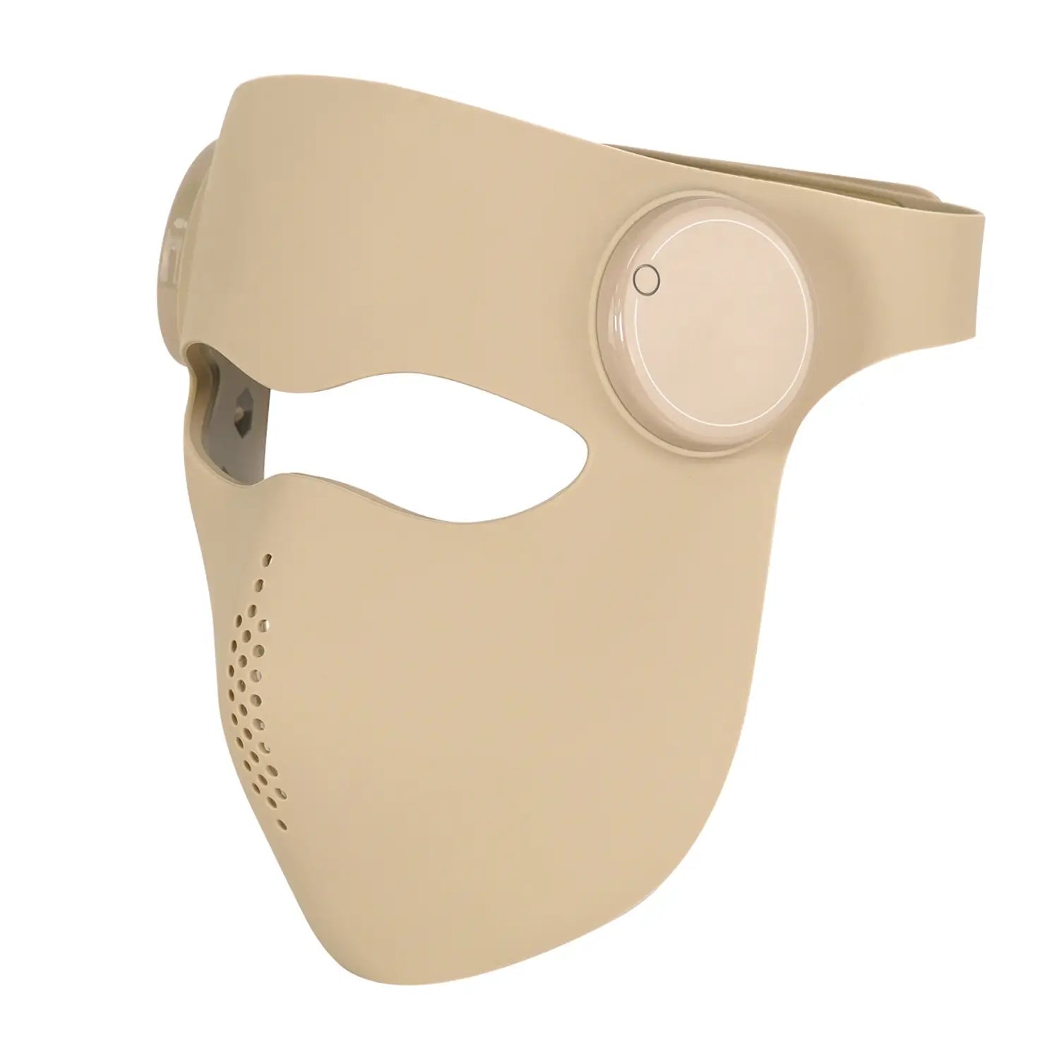
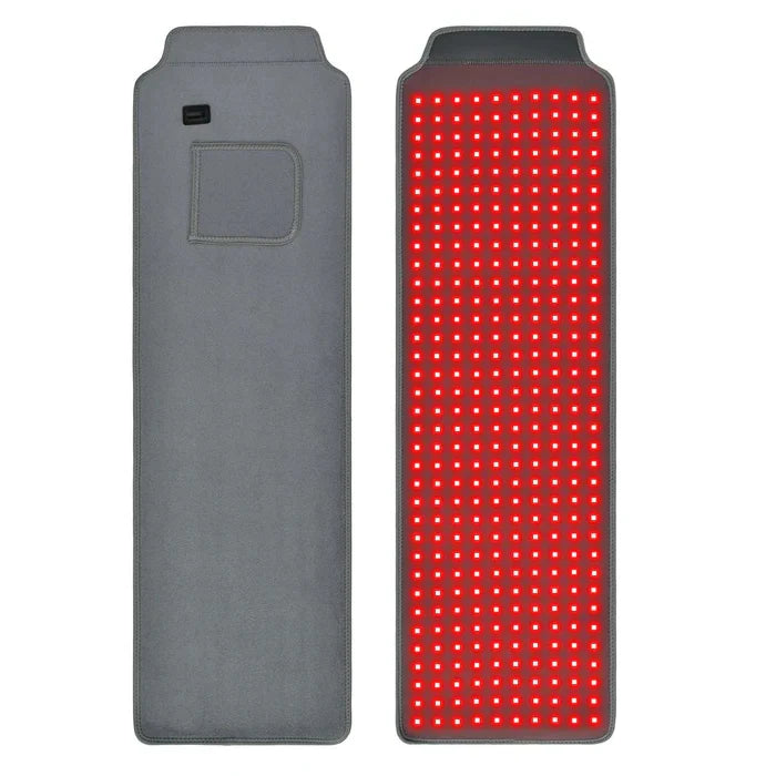
Fourth, match wavelength and target. The mesenchymal‑cell paper and the connective‑tissue review make it clear that 630–670 nm visible red and around 800–850 nm near‑infrared hit cells differently. For surface issues like fine lines, redness, and shallow scars, visible red between about 620 and 660 nm (common in facial panels) is well supported by dermatology data from sources such as Stanford Medicine and Cleveland Clinic. For deeper structures like joints, tendons, or fertility targets, near‑infrared around 800–830 nm, as used in some fertility case series, is more plausible.
Finally, respect your context. If you are on photosensitizing medications, have a history of skin cancer in the treatment area, or are dealing with an active malignancy, major centers such as MD Anderson and WebMD‑reviewed guidance recommend talking with a physician before adding aggressive light protocols. Gene‑expression modulation is powerful; you want it working for you, not blindly.
Pros, Cons, And Unknowns Of Targeting DNA Expression With Red Light
On the upside, red and near‑infrared photobiomodulation offer something rare in biology: a way to influence gene expression and repair pathways using an external physical input that, at appropriate doses, does not damage DNA.
By boosting mitochondrial output, calming excessive inflammation, and nudging key transcriptional hubs like TGF‑beta, Akt, STAT3, and GADD45A, red light can help tissues move from a “stuck” state into an active repair program. That shows up in the lab as altered transcriptional profiles in bone marrow stromal cells, fibroblasts, and osteoblasts. It shows up clinically as faster wound closure and less edema in surgical studies cited by dermatology groups, and as better collagen organization in both scar and anti‑aging research.
However, there are real limitations and open questions.
Many gene‑expression and animal studies have not yet been matched by large, rigorous human trials across all the conditions people market red light for. Stanford dermatology experts emphasize that evidence is strongest for hair growth and modest wrinkle reduction, while data for performance, sleep, and systemic disease remain weak or preliminary.
The dose problem is unresolved. The UC San Diego device patent, the MDPI connective‑tissue review, and the bone‑cell lethality at higher fluences all underscore that there is a narrow terrain between “no effect” and “too much,” and it depends on wavelength, cell type, and even the presence of photosensitizers. Consumer panels rarely provide fully transparent, independent dosimetry.
Long‑term gene‑expression consequences of chronic, whole‑body exposure are unknown. Most lab studies observe cells for hours to weeks after a single or short series of illuminations. We do not have multi‑year data on daily full‑body sessions in humans, particularly for people with pre‑existing genetic vulnerabilities.
And finally, cancer risk in complex scenarios remains an open field. Large reviews from MD Anderson, Cleveland Clinic, and WebMD agree there is no evidence that red light itself causes cancer and that it is fundamentally different from UV in this respect. But those same experts are cautious about using strong low‑level laser therapy over known tumors, precisely because shifting gene expression in already mutated cells could have unpredictable effects.
The takeaway is not to fear red light, but to treat it with the same respect you would any powerful biologic input.

Brief FAQ
Does red light change my DNA?
Red light in the 600–900 nm range, at doses used in clinics and well‑built home devices, does not change your DNA sequence and does not create UV‑type DNA lesions. What it does change is gene expression: which genes are turned on or off and how strongly. Studies in human stromal cells and dermal fibroblasts show hundreds to thousands of genes shifting expression after red‑light exposure, often through pathways like TGF‑beta, Akt, and GADD45A.
Can red light “turn on” longevity or repair genes?
To a degree, yes – but within biological limits. Laboratory work shows red light can increase expression of DNA repair mediators like GADD45A, shift Wnt signaling toward bone formation, and modulate antifibrotic and antioxidant pathways. Reviews of photobiomodulation also suggest activation of longevity‑related pathways such as AMPK, SIRT1, and PGC‑1alpha. In practice, this looks less like a magic “on switch” and more like gently biasing your cellular environment toward repair, provided you also support it with sleep, nutrition, and training.
Could red light make pre‑cancerous cells worse?
We simply do not know enough to give a blanket answer. There is no evidence that red light causes cancer in healthy tissues, and it does not carry the DNA‑damaging risk of UV. But in tissues that already harbor significant mutations, any intervention that boosts energy and alters signaling has a theoretical potential to either help immune clearance and repair or inadvertently support malignant survival. That is why major cancer centers recommend using red‑light and low‑level laser therapies under medical supervision in oncology settings and avoiding unsupervised high‑intensity treatment over known tumors.
Closing Thoughts
As a long‑time light‑therapy geek, what keeps me excited about red light is not the hype about “rewriting DNA,” but the subtler reality: a beam of non‑ionizing light can reach your cells, talk to their mitochondria, and nudge complex gene networks toward repair instead of breakdown, all without touching the genetic code itself.
Used thoughtfully – with respect for dose, wavelength, and your own biology – red and near‑infrared light become less a gadget and more a precision tool for steering DNA expression in the direction of healing and resilience. That is the kind of quiet, science‑backed leverage I want in any home wellness stack.
References
Karu T and colleagues on mitochondrial photoreceptors and ATP changes in low‑level light therapy.
Scientific Reports study on visible red and infrared light altering gene expression in human bone marrow stromal fibroblasts.
Mouse epidermis study on 670 nm red light modulating UV‑induced gene expression.
ScienceDirect article on visible red light enhancing GADD45A‑mediated DNA repair in normal human dermal fibroblasts.
NASA‑linked work on near‑infrared LED irradiation and wound‑healing gene expression in diabetic mice.
MDPI review on photobiomodulation, connective tissue cells, and repair processes in bone, cartilage, and tendon.
RNA‑seq analysis of red‑light phototherapy in human dermal fibroblasts with focus on fibrotic pathways.
Nature Communications article on visible light accelerating skin wound healing and modulating STAT3 signaling.
University of California San Diego patent on apparatus and methods for stimulating DNA repair using 630 nm red light.
Study on blue LED irradiation effects on proliferation and gene expression in cultured human keratinocytes.
New England Journal of Medicine work on UVB‑induced DNA damage and photolyase‑mediated repair in human skin.
Clinical overviews of red light therapy mechanisms, benefits, and safety from Cleveland Clinic, MD Anderson Cancer Center, Brown University, Stanford Medicine, and WebMD.
- https://techtransfer.universityofcalifornia.edu/NCD/30453.html
- http://ui.adsabs.harvard.edu/abs/2015SPIE.9309E..09B/abstract
- https://pmc.ncbi.nlm.nih.gov/articles/PMC26514/
- http://web.stanford.edu/group/barronlab/PubPdfs/2023/The%20anti-inflammatory%20effects%20of%20photobiomodulation%20are%20mediated%20by%20cytoki...ation..pdf
- https://www.ophth.wisc.edu/blog/2003/04/01/effect-of-nasa-light-emitting-diode-irradiation-on-molecular-changes-for-wound-healing-in-diabetic-mice/
- https://www.brownhealth.org/be-well/red-light-therapy-benefits-safety-and-things-know
- https://www.mdanderson.org/cancerwise/what-is-red-light-therapy.h00-159701490.html
- https://my.clevelandclinic.org/health/articles/22114-red-light-therapy
- https://www.rzhealth.org/the-recovery-room/what-is-red-light-therapy-health-benefits-explained/
- https://www.uclahealth.org/news/article/5-health-benefits-red-light-therapy