
Red light therapy is having a moment in the wellness world, but behind the glow panels and face masks there is a very real, very technical question: how does red light actually interact with the mechanics of collagen formation in your skin?
As someone who has spent years testing light devices, reading photobiomodulation papers late at night, and tweaking protocols down to the minute, I can tell you this: when you understand the collagen mechanics, the way you use red light changes. It becomes less “beauty gadget,” more “precision tool” for your extracellular matrix.
In this article, we will unpack both sides of the equation. First, how collagen molecules form, fold, and assemble. Then, how specific red and near‑infrared wavelengths alter the cellular and molecular environment that controls those mechanics, with evidence from controlled trials, in vitro studies, and collagen‑mimetic peptide research. Finally, we will turn all of that into practical guidance you can actually apply at home or in the clinic.
Collagen 101: From Triple Helix To Youthful Skin
Collagen is not just a “skin ingredient.” It is the most abundant protein in your body, accounting for roughly 30 percent of total protein and about 75 to 80 percent of your skin’s dry weight, according to clinical wellness sources that use whole‑body red light in practice. It forms the structural scaffolding for skin, muscles, bones, tendons, ligaments, blood vessels, and even the gut lining.
How A Collagen Molecule Is Built
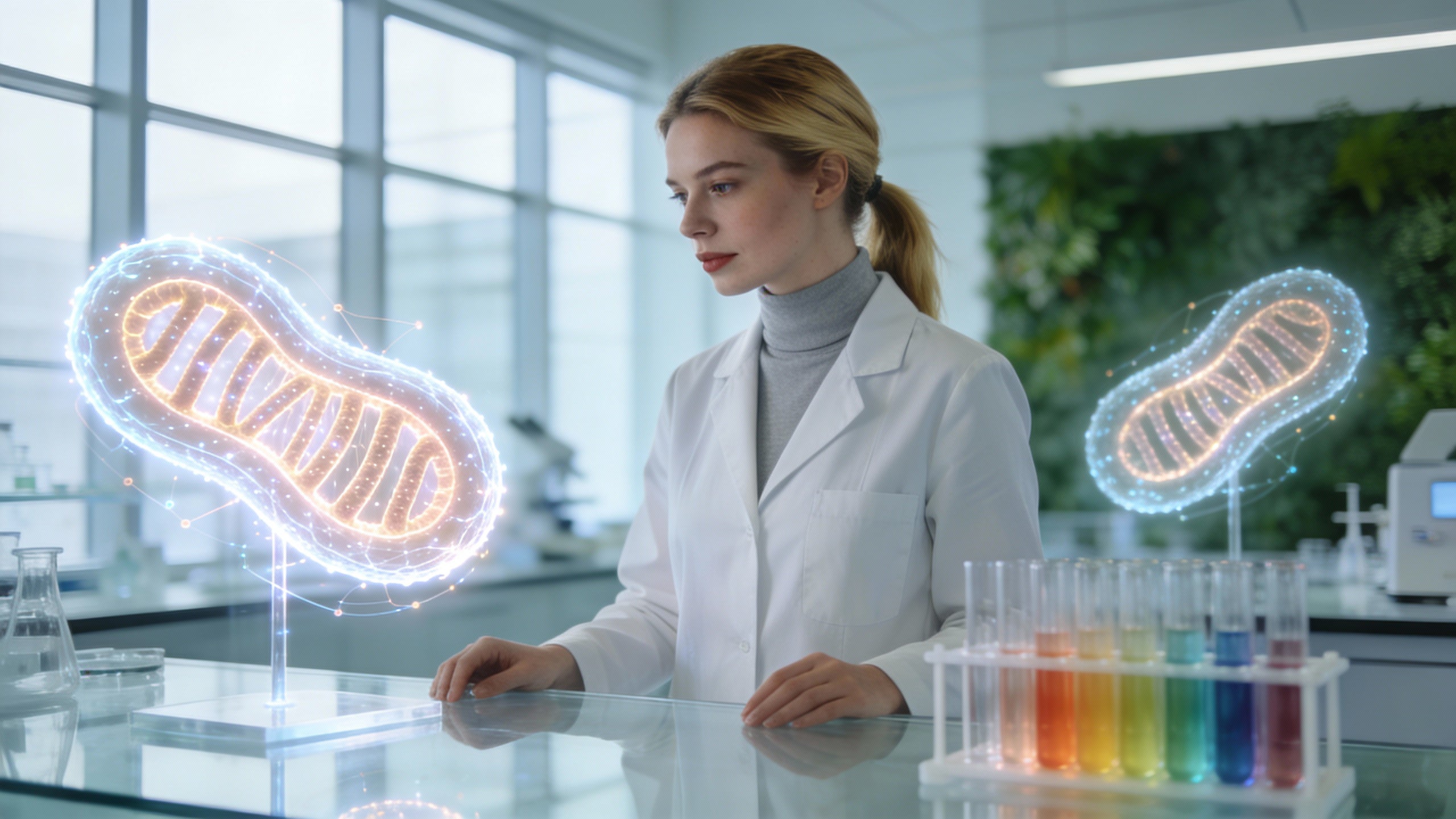
At the molecular level, collagen is built from three polypeptide chains wrapped around each other in a right‑handed triple helix. Each chain follows a repeating Gly–X–Y pattern, where glycine sits every third position and X and Y are often proline and hydroxyproline. Work from academic groups studying collagen‑mimetic peptides and triple‑helix stability describes this architecture as three left‑handed polyproline‑II‑like chains that twist into a single right‑handed superhelix.
Folding that triple helix is not trivial. Studies using model peptides show that collagen generally nucleates and folds from the C‑terminal end, then “zippers” toward the N‑terminus. When researchers remove or disrupt this C‑terminal nucleation domain, folding slows down and passes through more misaligned intermediates, even if the final helix is not dramatically less stable. This has been linked to real‑world collagen disorders, where mutations around the C‑terminus impair nucleation and early folding steps more than they change the final structure.
In other words, collagen formation is a kinetic problem as much as a structural one. The triple helix has to nucleate in the right place, in the right environment, and then assemble cleanly into higher‑order fibers and fibrils.
Environmental Control: pH, Charge, Hydrophobicity, And Metals
Collagen mechanics are also exquisitely sensitive to the local chemical environment. A dissertation using engineered collagen‑mimetic peptides demonstrated that:
Researchers added negatively charged carboxylate groups (labeled “PE”) to hydroxyproline and showed that a handful of these residues barely affected triple‑helix stability at acidic or neutral pH. However, when they increased to five to seven charged residues, the peptides could form a stable triple helix in acidic conditions but lost stability at neutral pH. As the number of negative charges rose, refolding became slower and thermal stability dropped because of electrostatic repulsion between strands.
In the same system, scientists introduced hydrophobic segments and metal‑binding residues. Hydrophobic “sticky ends” at the termini drove head‑to‑tail assembly into longer structures, while internal hydrophobic segments favored round, radially growing assemblies. When they added a metal‑binding amino acid and exposed the peptides to metal ions such as iron or gadolinium, the system formed diverse micro‑ and nanostructures depending on the ionic environment.
Taken together, these models show that collagen triple‑helix formation and higher‑order assembly are regulated by pH, electrostatic charge, hydrophobic patches, and local metal ions. Your dermal collagen is doing this dance all the time, in a living, dynamic extracellular matrix.
Why Collagen Declines With Age
Dermatology and photobiomodulation articles consistently point out that collagen production decreases with age, and one often‑cited estimate is roughly a 1 percent drop in collagen per year after about age 30. Combine two decades of that decline with chronic ultraviolet exposure, pollution, smoking, and hormonal changes such as lower estrogen, and you get thinner, less elastic skin with more wrinkles and sagging.
If collagen is about 75 to 80 percent of your skin’s dry weight, even a modest percentage decrease in production and an increase in breakdown change the mechanical behavior of the entire matrix. From a mechanics perspective, you are losing both load‑bearing capacity and optimal triple‑helix packing.
Now the key question for a light‑therapy geek is: where does red light fit into this molecular story?

Photobiomodulation: What Red Light Actually Does To Skin Cells
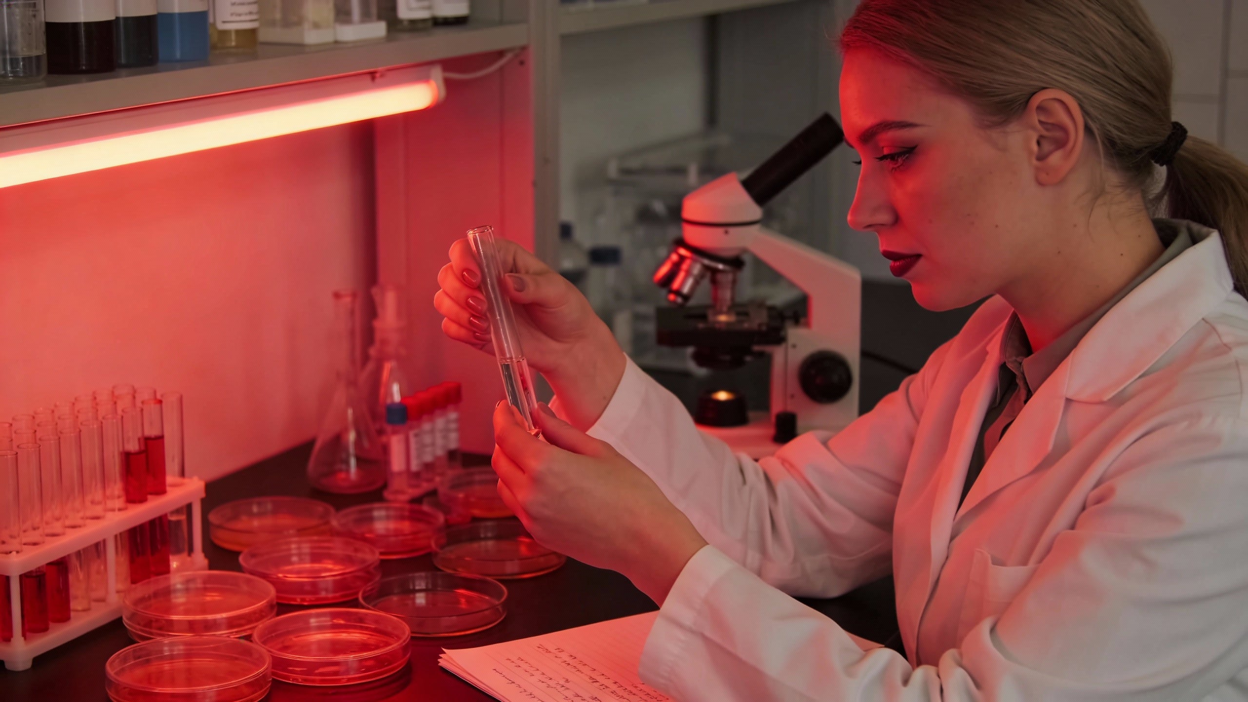
Red light therapy, also known as low‑level light therapy or photobiomodulation, uses specific red and near‑infrared wavelengths (roughly 600 to 1,100 nanometers) at low power to modulate biology without heating or ablating tissue. Medical centers, dermatology clinics, and academic reviews describe this as “atraumatic” and “non‑ablative” because, unlike lasers that deliberately damage tissue to trigger repair, red light directly stimulates regenerative processes.
Mitochondria As The First Target
Across clinical reviews, Cleveland Clinic–style patient guides, and mechanistic studies, the core mechanism is consistent. Photons in the red and near‑infrared range are absorbed by mitochondrial chromophores, especially cytochrome c oxidase. This leads to increased mitochondrial membrane potential, higher ATP output, and downstream shifts in gene expression.
A detailed ex vivo and cell‑culture study using a combination of 640 nanometer red and 830 nanometer near‑infrared light at very low power (about 0.5 milliwatt per square centimeter for 10 minutes, or 0.3 joule per square centimeter) found several key changes in human dermal fibroblasts and skin explants. The treatment upregulated genes for type I and type III collagen (COL1A1 and COL3A1), elastin (ELN), and lysyl oxidase‑like 1 (LOXL1), which is involved in crosslinking elastic fibers. It also increased type I procollagen and elastin proteins and raised ATP levels.
In other words, red plus near‑infrared light does not just turn on collagen genes; it appears to support the energy budget and maturation of collagen and elastin fibers.
Anti‑Inflammatory And Vascular Effects
Stanford dermatology and other clinical overviews emphasize another layer of biology: red light induces vasodilation and improves microcirculation. Wider blood vessels mean better delivery of oxygen and nutrients to fibroblasts and other skin cells.
At the same time, multiple sources describe red light as anti‑inflammatory and anti‑oxidative. It reduces pro‑inflammatory markers and oxidative stress in various tissues. Clinics that use whole‑body panels report benefits in conditions ranging from acne and rosacea to tendonitis, bursitis, and arthritis, with inflammation and pain reduction as a unifying theme.
This matters for collagen mechanics because inflammatory and oxidative environments tend to acidify tissue, alter ionic strength, and generate reactive species that can damage proteins or disrupt folding. By calming that environment and improving circulation, red light plausibly shifts the extracellular conditions closer to the pH and ionic ranges where collagen peptides fold and assemble efficiently in the lab.
Wavelengths, Dose, And The Arndt–Schulz Principle
Not all light is created equal, and the dose matters. A controlled trial of a commercial red‑light mask, conducted under Good Clinical Practice standards, used cold red LEDs at 630 nanometers (plus or minus 10 nanometers) with a power density of about 21.7 milliwatts per square centimeter. Each 12‑minute session delivered around 15.6 joules per square centimeter, applied twice weekly for three months to volunteers aged roughly 45 to 70.
In parallel, a large randomized trial of full‑body light cabins used gas‑discharge lamps with either a narrowband red spectrum (611 to 650 nanometers) or a broader 570 to 850 nanometer band, normalized to about 9 joules per square centimeter in the red window. Session durations ranged from 12 to 25 minutes, given twice weekly for 30 sessions.
The mask study explicitly referenced the Arndt–Schulz law: too little light and you get no biological response; too much and you can actually inhibit or blunt the effect. That shows up in the numbers. The in vitro collagen‑and‑elastin study used only 0.3 joule per square centimeter per day to trigger gene changes, while the mask used roughly 15.6 joules per square centimeter twice a week. The full‑body cabins sat in between on a per‑session basis but treated a much larger area.
This is one reason cookie‑cutter dosing rules are misleading. The right dose domain is determined by wavelength, irradiance, session length, and how often you repeat the exposure.
For a simple thought experiment, consider the mask protocol. Two sessions per week for twelve weeks at 15.6 joules per square centimeter per session yields a cumulative exposure around 375 joules per square centimeter over three months for the treated areas. The collagen‑mimetic peptides used to map folding mechanics in the lab respond to differences of just a few joules per square centimeter in terms of heating or stress. That gives you a sense of how much integrated energy you are introducing into the system over time, even at “low level” settings.
How Red Light Interfaces With Collagen Formation Mechanics
Now we can connect the dots. Collagen mechanics come down to fibroblast behavior, triple‑helix folding and refolding, and higher‑order assembly in a pH‑ and charge‑sensitive matrix. Red light reshapes the inputs to that system.
From Mitochondria To Triple‑Helix Output
The 640 plus 830 nanometer in vitro work is a useful starting point. By showing increased expression of type I and type III collagen, elastin, and LOXL1, along with higher ATP, it demonstrates that red and near‑infrared light can simultaneously:
Increase the number of collagen chains being synthesized by fibroblasts.
Provide more energy for quality‑controlled folding and post‑translational processing.
Support maturation and crosslinking of the elastic fiber network.
That aligns well with clinical data. In the large randomized trial with 136 volunteers, subjects receiving either narrowband red light (611 to 650 nanometers) or broader 570 to 850 nanometer light twice weekly for 30 sessions showed significant increases in intradermal collagen density by high‑frequency ultrasound. The collagen intensity score rose by about six units on average in both treatment groups, while controls showed no meaningful change.
At the same time, profilometry of the periorbital area showed reduced skin roughness in both light groups and a worsening of roughness in controls. Blinded physicians rating standardized photographs judged wrinkles improved in roughly 69 percent of the narrowband red group and 75 percent of the polychromatic group, compared with about 4 percent of controls.
From a mechanics viewpoint, that combination of higher collagen density and lower surface roughness suggests not just more collagen, but better organized collagen that supports a smoother, more elastic surface.
pH, Charge, And The Microenvironment
The peptide studies on pH‑responsive collagen give us a sharper way to think about red light’s anti‑inflammatory benefit. When peptides carry more negatively charged residues, their triple helices fold more slowly and are less thermally stable at neutral pH, although they can remain stable under acidic conditions. Change the pH or ionic balance and you change the folding landscape.
Inflamed, poorly perfused tissue is typically more acidic and more ionically imbalanced than healthy tissue. Red light’s ability to increase blood flow and reduce inflammatory markers, as observed in both clinical practice and mechanistic reviews, plausibly shifts those microenvironmental variables in the direction that favors faster, cleaner triple‑helix folding and refolding. That is a mechanistic hypothesis rather than a fully proven pathway, but it is grounded in how charged collagen peptides behave in controlled experiments.
When you add in the hydrophobic and metal‑binding findings, the picture gets richer. Hydrophobic segments at the ends of collagen peptides can create “sticky ends” that drive head‑to‑tail assembly, while internal hydrophobic segments promote radial growth. Metal‑binding residues coordinate ions like iron and gadolinium into diverse nanostructures. Real dermal collagen is not engineered with those exact designs, but it does live in a matrix rich in lipids, minerals, and metal ions. Better circulation and lower oxidative stress should help maintain those gradients in a range that supports healthy fiber assembly rather than disorganized, scar‑like structures.
Nucleation, Folding Kinetics, And Why Results Take Weeks
The triple‑helix folding work on C‑terminal nucleation domains also helps explain why clinical improvements from red light are gradual and cumulative rather than instant.
In the nucleation‑domain studies, removing the stable C‑terminal segment forced peptides to find alternative, less efficient nucleation pathways. Folding became slower and more complex, with misaligned intermediates. Translating that to skin, your fibroblasts are constantly synthesizing procollagen, folding it into triple helices, and secreting it into the matrix, where it assembles into fibrils. Even if red light doubles the rate of collagen chain synthesis at the gene and protein level, the actual assembly and remodeling of the dermis is bottlenecked by folding kinetics and matrix turnover.
Look at the timelines from human trials. The red‑light mask study measured objective and clinical endpoints at about one, two, and three months. Improvements in crow’s feet wrinkle depth, facial sagging, firmness, elasticity, dermal density, skin roughness, complexion homogeneity, and pores all increased progressively over the three‑month period rather than appearing at the first check‑in. Importantly, benefits persisted for roughly one month after stopping treatment before gradually tapering, suggesting genuine structural remodeling rather than fleeting vasodilation.
The full‑body cabin study used 30 sessions over several months. Subjective skin feeling and complexion improved significantly over that period, collagen density increased, roughness decreased, and wrinkle grades improved compared with untreated controls. A subset followed for about six months after completing 30 sessions still showed better scores than at baseline, even though some of the gains had attenuated.
In my own optimization experiments, I treat red light more like strength training for fibroblasts than like makeup. You do not build new tensile structure in a single session. You apply the stimulus repeatedly, give the system time to fold and assemble triple helices, and accept that the decay after stopping will also be gradual.
Quantitative Snapshot: How Protocols Compare
Here is how several evidence‑based protocols line up when you think in terms of collagen mechanics.
Scenario |
Wavelengths |
Approximate dose per session |
Frequency and duration |
Key collagen‑related outcomes |
At‑home facial mask trial |
630 ± 10 nm red |
About 21.7 mW/cm²; 15.6 J/cm² in 12 min |
Twice weekly for 3 months |
Increased dermal density, firmness, elasticity; reduced wrinkles and roughness; more even tone; benefits lasting about 1 month post‑treatment |
Full‑body cabin randomized trial |
611–650 nm red, or 570–850 nm |
About 8.5–9.6 J/cm² in 12–25 min for red band |
Twice weekly for 30 sessions |
Higher intradermal collagen density; smoother skin; majority with wrinkle improvement vs minimal change or worsening in controls |
In vitro fibroblast and skin explant study |
640 nm red + 830 nm near‑IR |
0.3 J/cm² in 10 min at 0.5 mW/cm² |
Once daily in lab setting |
Upregulated collagen and elastin genes; more type I procollagen; stronger elastic fiber formation and crosslinking; higher ATP |
What jumps out is the order‑of‑magnitude difference in dose between the lab study and the clinical mask and cabin trials, yet all of them show collagen‑relevant changes. That again reflects the Arndt–Schulz principle: there is a therapeutic window, and you can hit it with a range of dose patterns as long as irradiance and timing are tuned.

Practical Guidance: Using Red Light To Support Collagen Mechanics
All of this theory is useless if it does not translate to decisions you can make about your own routine. Here is how I think about designing collagen‑centric photobiomodulation protocols that respect the underlying mechanics and the actual evidence.
Choosing A Device And Wavelength Strategy
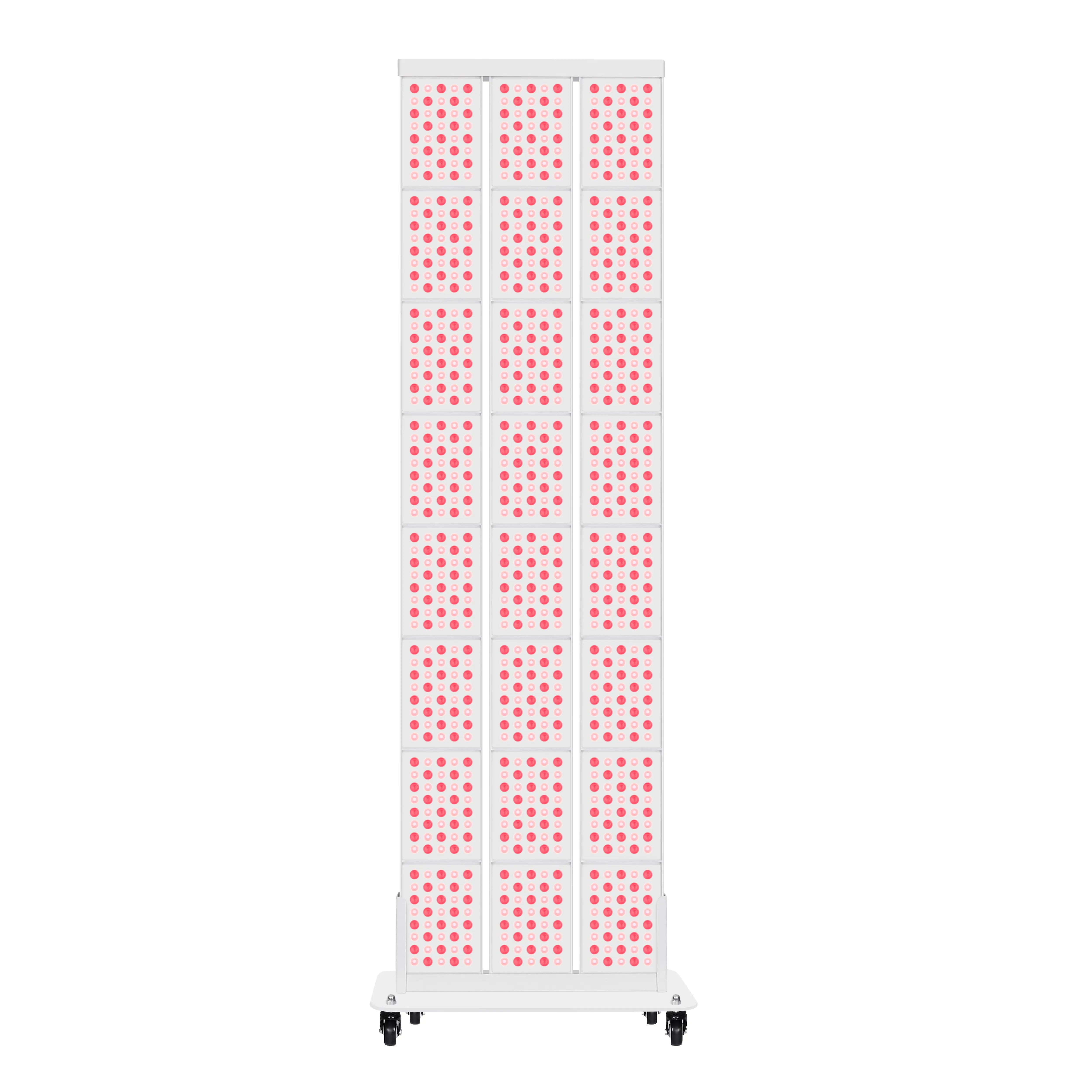
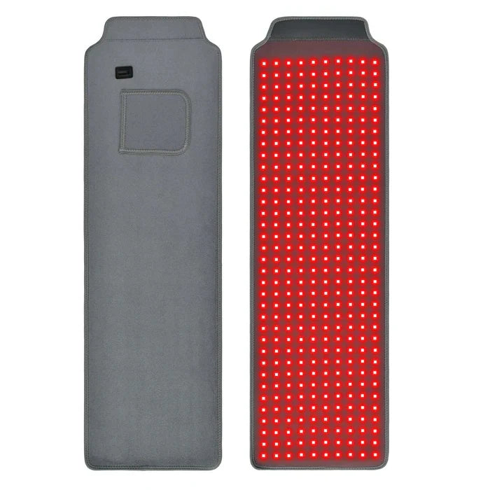
Multiple clinical and safety reviews note that both visible red and near‑infrared light have roles. Visible red in the 630 to 660 nanometer range penetrates into the dermis and directly targets fibroblasts and superficial vascular networks. Near‑infrared, around 800 to 880 nanometers, dives deeper into subdermal tissues and inflammatory pathways.
The fact that the full‑body trial found no major advantage of the broader 570 to 850 nanometer spectrum over the narrower 611 to 650 nanometer spectrum for collagen density tells us something important. You do not necessarily need an exotic multi‑band device to affect collagen. Well‑designed red‑dominant systems at appropriate power densities can be enough.
When I evaluate a device for collagen work, I look for three things that are backed by published protocols. First, does it specify peak wavelength in a range similar to 630 nanometers or 611 to 650 nanometers, or pair that kind of red light with a near‑infrared band around 830 nanometers. Second, does it publish irradiance numbers so I can estimate whether a 10 to 20 minute session will land near the 10 to 20 joule per square centimeter range used in human face and full‑body trials. Third, is it designed for consistent, repeatable exposures over weeks, not just short “spa novelty” sessions.
Dermatology and academic sources also note that at‑home devices are usually lower power than clinic‑grade systems. That is not inherently bad; it just means you must lean more on consistency and proper positioning and less on any single heroic dose.
Building A Collagen‑Friendly Protocol At Home
Most reputable medical and wellness organizations converge on similar practical advice, even if the exact parameters differ.
Sessions of roughly 10 to 20 minutes, several times per week, are typical in both consumer guides and clinical settings. The mask trial shows that twice‑weekly use for three months can produce measurable changes in wrinkles, sagging, firmness, dermal density, and sebum output. Physical therapy and dermatology clinics often recommend an initial phase of more frequent sessions over about six to twelve weeks, followed by a maintenance phase once weekly or every other week.
That lines up nicely with what we know about collagen mechanics. A sustained stimulus over at least one to three months allows fibroblasts to increase production, triple helices to fold and assemble, and your dermis to remodel. Once you have that new structure, less frequent “reminders” can maintain it, though stopping completely will gradually allow the system to drift back toward baseline.
From an execution standpoint, several clinic protocols and whole‑body practices emphasize simple but important details. Go into a session with clean, dry skin and no sunscreen or makeup over the treated areas. Use eye protection, especially for high‑intensity panels or full‑body cabins. Stay well hydrated, and avoid alcohol immediately around sessions. Some providers also recommend not loading up on saturated fats right before treatment, aiming to keep blood viscosity and circulation optimized.
I would add a mechanics‑driven tip: protect your recovery window. Just as you would not follow a heavy strength workout with nightly all‑nighters and junk food and expect peak muscle gains, you should not treat red light sessions as a license to ignore sleep, basic nutrition, or photoprotection. Several dermatology sources stress pairing red light with a solid skincare routine that includes daily sunscreen and gentle topicals rather than harsh peels or excessive actives.
Pros, Cons, And Safety Boundaries
Compared with lasers, chemical peels, and injectables, red light therapy has clear advantages and real limitations.
On the plus side, it is non‑invasive, non‑UV, and generally well tolerated. Trials of full‑body cabins and at‑home masks reported no serious adverse events and very few discontinuations. Minor side effects, when they occurred, were limited to temporary redness, warmth, or light sensitivity. In the big cabin study, a pre‑existing facial telangiectasia became more visible in one participant and an old knee scar reddened briefly in another, both manageable events.
Medical groups such as Cleveland Clinic, the American Academy of Dermatology, and academic centers highlight that red light is non‑ionizing, which means it does not have the energy to damage DNA the way ultraviolet light can. Dermatology associations still urge eye protection and caution users that misuse of devices (too high an intensity, too long, or too close to the skin) can cause irritation or, in rare cases, burns.
On the con side, experts are blunt about the limitations. Evidence for wrinkle reduction and modest collagen improvements is solid but not miraculous. Parameters such as exact wavelength, dose, and frequency are not standardized across devices, which makes direct comparison difficult. Studies for systemic claims, like boosting athletic performance, treating dementia, or radically improving chronic pain, are early and inconsistent. Several major health systems stress that there is no good evidence for red light as a treatment for weight loss, cellulite, cancer, or mental health conditions such as depression.
There are also population‑specific considerations. Some dermatology resources note that people with darker skin may be more prone to temporary pigmentation shifts with visible light treatments, which argues for professional supervision and conservative dosing. Individuals on photosensitizing medications, those with active skin cancer or suspicious lesions, and pregnant patients are routinely advised to clear red light therapy with a physician before starting.
The bottom line from a mechanics‑focused perspective is that red light is a low‑risk way to nudge your collagen system toward more favorable energy and signaling states, not a license to bypass medical care or chase extreme, unproven outcomes.
Collagen Biohacking With Integrity
My own philosophy as a “light therapy geek” and long‑time wellness optimizer is simple: respect the biology, respect the data, and then experiment within that frame.
On the biology side, collagen formation is about more than just turning on a gene. It requires proper nucleation of the triple helix, balanced charge and pH, adequate energy and co‑factors for folding, and space in the matrix for fibers to assemble and remodel. Red light helps by raising ATP, activating fibroblasts, smoothing out inflammatory noise, and improving microcirculation, all of which support those mechanics.
On the data side, we now have controlled trials showing that realistic red‑light doses can increase dermal collagen density, reduce wrinkle depth, improve elasticity, and smooth roughness over multi‑month time frames. We have peptide and in vitro studies showing direct effects on collagen and elastin gene expression and structural maturation. And we have safety data indicating a favorable profile when treatments are performed thoughtfully.
Within that frame, your job as an informed user is to choose devices and protocols that echo what has been shown to work. That means favoring known wavelengths, doses in the ranges used in masks and cabins, and schedules that build up stimulus over weeks, not days. It means combining light with lifestyle, not treating it as a substitute for sleep, nutrition, or photoprotection. And it means staying honest about what is proven, what is promising, and what is still speculative.
Used this way, red light is not just a cosmetic trick. It is a targeted way to influence the mechanics of one of your body’s most fundamental structural systems: the collagen triple helix and its living matrix. When you align your protocols with those mechanics, you give your skin a real chance to remodel from the inside out.

References
- https://jdc.jefferson.edu/cgi/viewcontent.cgi?article=1146&context=orthofp
- https://pubmed.ncbi.nlm.nih.gov/33594706/
- https://www.princeton.edu/~persikov/project-collagen.html
- https://ui.adsabs.harvard.edu/abs/2017nsf....1709631H/abstract
- https://docs.lib.purdue.edu/dissertations/AAI3413749/
- http://labs.cas.usf.edu/cbb/Papers/Cgen_final_52316_printed2pdf.pdf
- https://digitalcollections.ohsu.edu/record/2580/files/3323_etd.pdf;
- https://dspace.mit.edu/bitstream/handle/1721.1/110088/Molecular%20level%20detection.pdf?sequence=1&isAllowed=y
- https://scholarship.libraries.rutgers.edu/esploro/outputs/journalArticle/Transformation-of-the-mechanism-of-triple-helix/991031665225004646
- https://med.stanford.edu/news/insights/2025/02/red-light-therapy-skin-hair-medical-clinics.html