

Why Membrane Potential Is The “Missing Variable” In Light Therapy
If you spend any time in biohacking or performance circles, you hear the same red light talking points on repeat: more ATP, less inflammation, better skin. Those are real themes in the literature, but they all rest on something much more fundamental and much less discussed: the electrical potential across your cell membranes.
Your cells live in an electrical universe. Every neuron firing, every muscle contraction, every immune response depends on a voltage difference between the inside and outside of membranes. That is true for the outer cell membrane and for the inner mitochondrial membrane that drives ATP synthesis.
Several of the strongest red and near‑infrared light studies do not just talk about “energy” in the abstract. They explicitly track mitochondrial membrane potential, proton gradients, and redox state. A vision‑focused review from Mitohealth, for example, emphasizes that the retina is the most energy‑hungry tissue in the body and that low‑dose red and near‑infrared light can raise mitochondrial membrane potential and ATP in retinal cells while reducing inflammation. A Nature review on near‑infrared countermeasures for spaceflight‑associated neuro‑ocular syndrome highlights the same mechanism in stressed astronaut eyes: red and near‑infrared light improves mitochondrial inner membrane potential, boosts ATP, and dampens oxidative stress.
At a practical level, membrane potential is the voltage that lets mitochondria run their turbines and lets your ion pumps keep sodium, potassium, and calcium exactly where they belong. When that voltage sags, you feel it as fatigue, brain fog, slow recovery, and cellular “under‑recovery.” Red light therapy, properly dosed, is one of the few tools that can nudge that voltage without drugs.
In the rest of this article, I will walk through how that works, what the evidence actually shows, how to think about dosing at home versus in a clinic, and where the hype clearly outruns the data.

Photobiomodulation 101: What Red Light Really Does To Cells
Scientists usually call red light therapy photobiomodulation, low‑level light therapy, or simply PBM. Across sources such as Cleveland Clinic, US military pain research, and multiple PubMed‑indexed reviews, the core definition is consistent: low‑power red and near‑infrared light between roughly 600 and 1,000 nanometers, delivered by LEDs or non‑thermal lasers, used to modulate biology without heating or burning tissue.
At these wavelengths, the main “antenna” is not your skin pigment. It is the mitochondrial enzyme cytochrome c oxidase, the last complex in the electron transport chain. A series of mechanistic papers summarized in journals like Photochemistry and Photobiology and IUBMB Life converge on a model where red and near‑infrared photons are absorbed by cytochrome c oxidase and other chromophores. That absorption speeds electron flow down the respiratory chain, pumps more protons across the inner mitochondrial membrane, and thereby strengthens the electrical and chemical gradient that powers ATP synthase.
A large immunomodulation review in PubMed Central reports that this mitochondrial stimulation can increase ATP production by up to about 70 percent in some cell types. A regenerative medicine article notes that 670 nanometer red light has been linked to roughly a 25 percent rise in ATP in stressed cells. A fibroblast study comparing 636 nanometer red light to 825 nanometer near‑infrared light found that the near‑infrared exposure produced roughly double the ROS and nearly double the ATP output of red light across the doses tested.
Membrane potential and ATP are tightly coupled: more electron flow and proton pumping strengthen the inner membrane potential, and that higher potential, in turn, makes ATP synthase more efficient.
However, photobiomodulation is not just an energy booster. It is a redox modulator. The immunomodulation review and redox‑focused work in journals like Current Biology and Photomedicine show a biphasic response in reactive oxygen species, meaning dose matters tremendously. Low doses around 5 to 10 joules per square centimeter tend to reduce excessive ROS in already stressed tissues, acting like an antioxidant nudge. Higher doses, in the 20 to 50 joules per square centimeter range, transiently increase ROS in otherwise healthy cells, which acts as a growth and repair signal.
That same review shows that these shifts in ROS and ATP ripple into major signaling pathways. Nuclear factor kappa B (NF‑kappaB), a master regulator of inflammatory gene expression, can be dialed down in inflamed tissues by roughly 30 percent, lowering pro‑inflammatory cytokines. Mitogen‑activated protein kinase (MAPK) cascades, including ERK and p38, are modulated in ways that promote survival and fine‑tune cytokine profiles.
In other words, red light therapy is not magic. It is a controlled way to tweak the mitochondrial membrane potential and redox balance that sit upstream of inflammation, repair, and, by extension, cell membrane potential and excitability.
Evidence That Red Light Shifts Mitochondrial Membrane Potential
The Retina: High‑Voltage Tissue And Low‑Dose Red Light
If you want to see membrane potential effects clearly, look at tissues that live on the edge energetically. The retina is an ideal case study. It is packed with mitochondria and relies on very high mitochondrial membrane potential to keep photoreceptors and retinal ganglion cells firing cleanly.
The Mitohealth review on red light and vision points out that gentle, low‑dose red or near‑infrared light in the 600 to 1,000 nanometer range can raise mitochondrial membrane potential and ATP production in retinal cells while easing inflammation. Pediatric clinics already use tabletop red light devices several times per week to slow myopia progression, based on trials showing reduced myopia onset and slower progression compared with usual care. Those trials are not framed in “biohacker” language; they are built around membrane potential, ATP, and tissue resilience.
A Nature paper on near‑infrared or red light as a countermeasure for astronaut neuro‑ocular issues goes further. It describes how 670 nanometer light improves contrast sensitivity and scotopic thresholds in older adults with just three minutes of exposure within a defined testing window. In aging eyes, these improvements correlate with enhanced mitochondrial inner membrane potential and reduced oxidative stress.
A related Nature study on longer wavelengths in sunlight shows that 850 nanometer light can transmit across the human body and improve visual function even when applied away from the eyes, indicating a systemic mitochondrial effect. The authors tie this to improved mitochondrial membrane potential and ATP production in multiple tissues, with multi‑day benefits after very brief exposures.
Taken together, these ophthalmic and systemic data show a consistent pattern. When you deliver carefully controlled doses of deep red or near‑infrared light, you can measurably improve mitochondrial membrane potential in energy‑hungry tissues and see functional gains in vision.
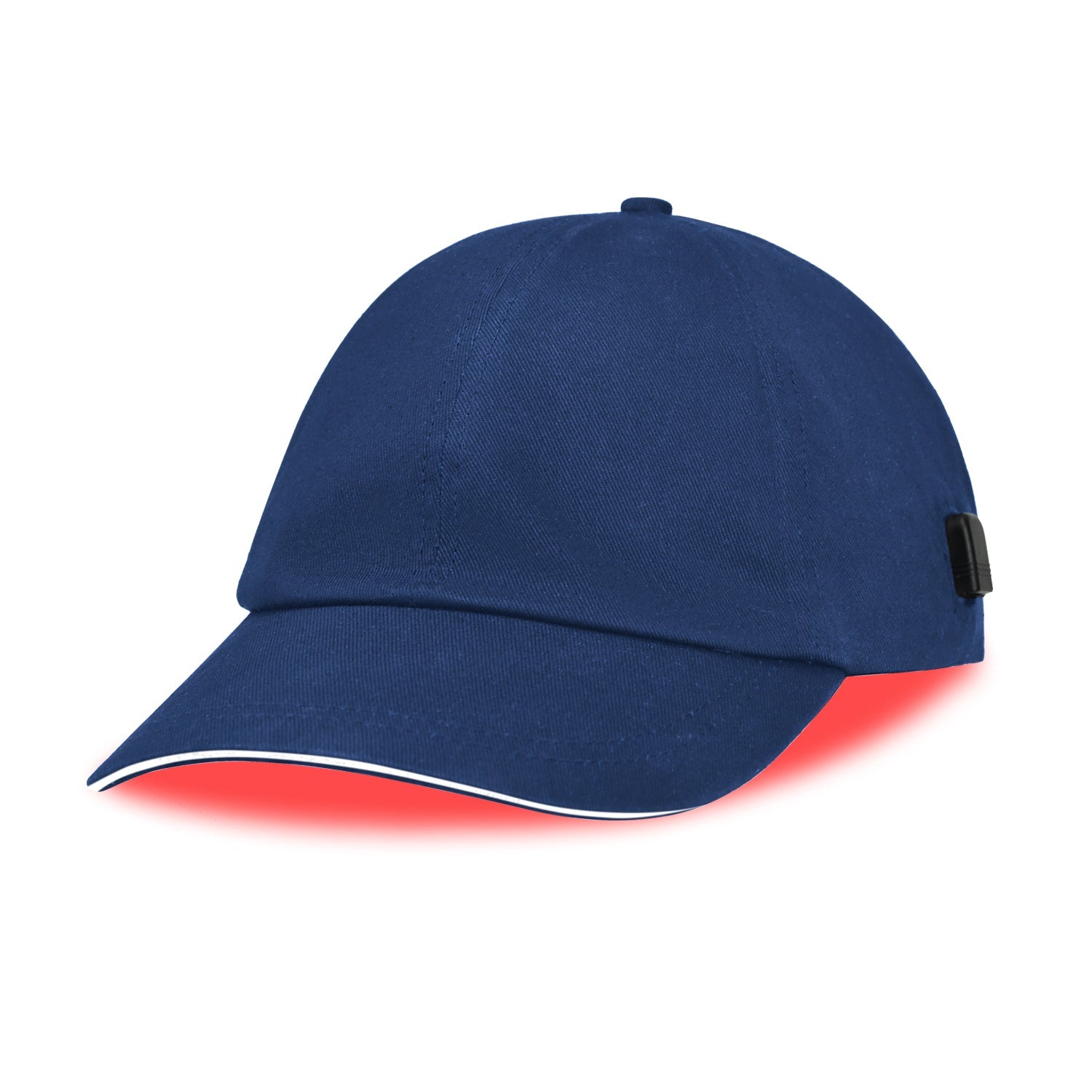
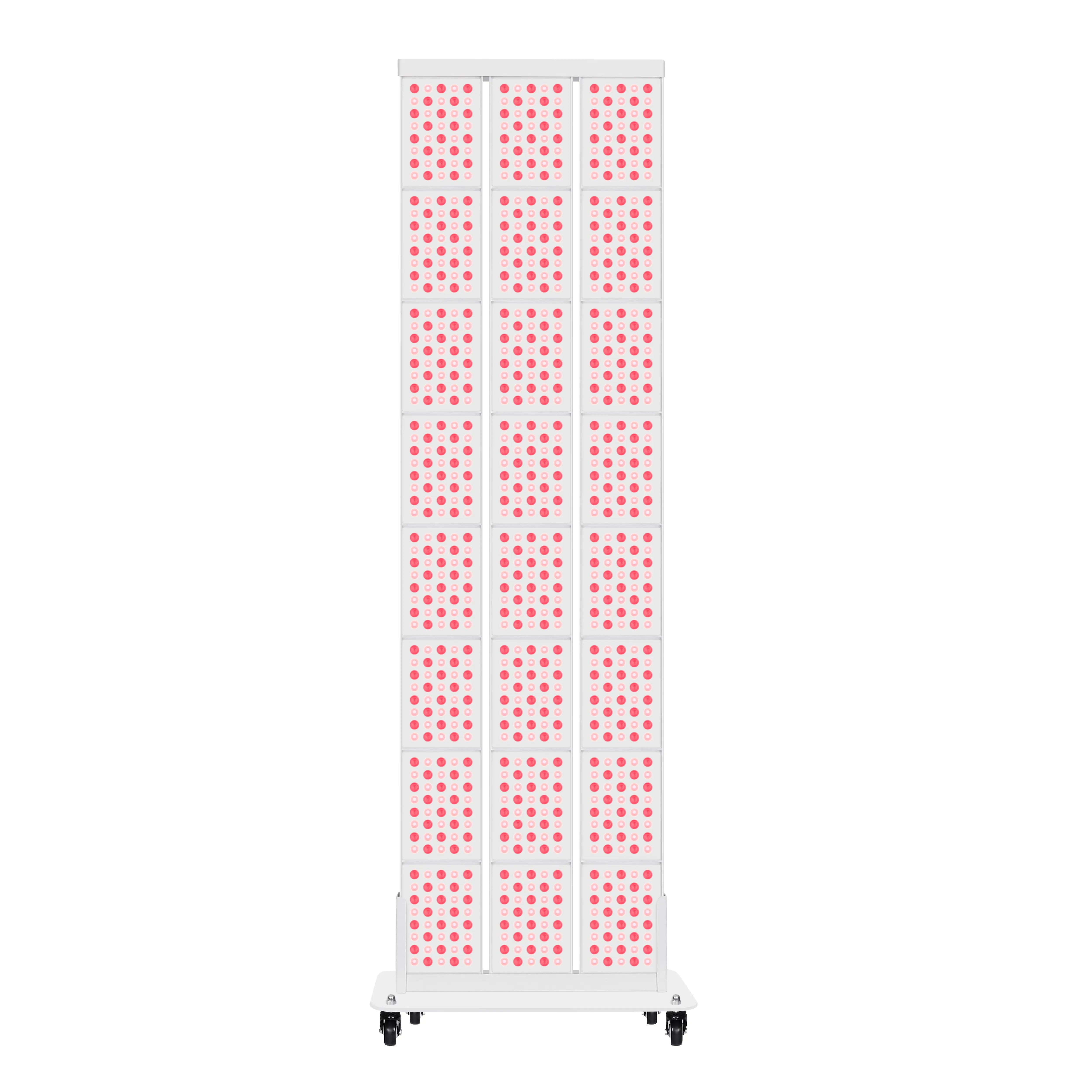
As a concrete example, consider a clinic that uses a panel delivering about 10 milliwatts per square centimeter of 670 nanometer light to the face. A ten‑minute exposure translates into roughly 6 joules per square centimeter at the skin surface, which sits right in the low‑dose window described in several immunomodulation and fibroblast studies. That is the same general dose range where retinal and brain benefits have been seen.
Fibroblasts And Skin: ROS, ATP, And Electrical Readiness
Skin is another useful test bed because it is accessible and easy to biopsy. An article in Scientific Reports looked at human dermal fibroblasts exposed to 636 nanometer red light versus 825 nanometer near‑infrared light across fluences from 5 to 25 joules per square centimeter. The near‑infrared light consistently generated roughly twice as much ROS as red light and drove about double the metabolic activity and ATP as measured in those cells, while maintaining acceptable viability.
That pattern matters for membrane potential. Mitochondrial ATP production is directly tied to the proton gradient and voltage across the inner mitochondrial membrane. When fibroblasts show higher ATP and controlled ROS without cell death, it implies that the mitochondrial machinery is running “hotter” without burning out, which is exactly the type of state you want if you are trying to speed wound healing or collagen synthesis.
Cleveland Clinic’s overview on red light therapy notes that most dermatologic uses under study, from wrinkles and age spots to scars and inflammatory dermatoses, are hypothesized to work through this mitochondrial pathway: more ATP, more collagen, better blood flow, and lower inflammatory markers. Hundreds of dermatology studies, summarized in outlets like the Journal of Clinical and Aesthetic Dermatology and Stanford’s dermatology insights, show modest but real improvements in wrinkles and skin thinning when red light is used correctly. Those improvements track with increased collagen density and, beneath the surface, more robust mitochondrial membrane potential in fibroblasts and keratinocytes.
Immune Cells: Membrane Potential, Inflammation, And Autoimmunity
Immunology may feel far from membrane potential, but immune cells are as dependent on mitochondrial voltage as neurons are. The comprehensive immunomodulation review in PubMed Central synthesizes clinical and mechanistic work on photobiomodulation across autoimmune and inflammatory diseases.
At the cellular level, red and near‑infrared light between 600 and 1,000 nanometers increase cytochrome c oxidase activity, proton gradients, and ATP, just as in retina and skin. In immune cells specifically, this results in enhanced phagocytosis, controlled ROS, and different cytokine profiles. Macrophages are nudged toward an M2 pro‑repair phenotype, with increased expression of markers like CD206 and Arginase‑1 and higher secretion of anti‑inflammatory cytokines such as IL‑10 and TGF‑beta.
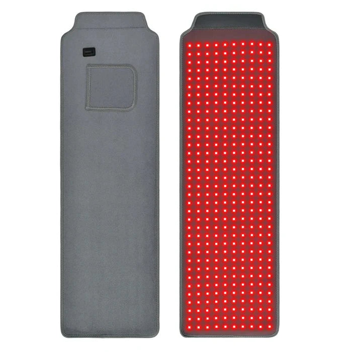
Clinically, the same review describes rheumatoid arthritis patients treated with 810 nanometer light at 10 joules per square centimeter three times per week for twelve weeks. Compared to controls, they experienced about a 45 percent reduction in pain scores, a 38 percent improvement in morning stiffness, and around a 30 percent drop in pro‑inflammatory cytokines, without significant adverse effects. Chronic wound patients treated with 660 nanometer light at 4 joules per square centimeter daily saw wound closure rates roughly 65 percent faster, along with increased M2 macrophage polarization and improved tissue regeneration.
All of these changes require intact membrane potentials across mitochondrial and plasma membranes in macrophages, T cells, and other immune cells. By stabilizing mitochondrial membrane potential and moderating ROS, red light appears to keep immune cells in a more responsive, less chronically inflamed state.
Aging Flies, ATP, And Systemic Voltage
For a whole‑organism view, consider a study in Scientific Reports on aged fruit flies exposed to 670 nanometer near‑infrared light for twenty minutes per day. After seven weeks, flies receiving light had about 80 percent higher whole‑body ATP compared with controls. Systemic inflammation markers were lower by about 15 percent, and mobility tests showed that treated flies climbed roughly twice as far and as fast.
Interestingly, survival curves shifted as well. From week three onward, treated flies outlived controls, with about 10 percent more survivors at week four and over 100 percent more survivors by week eight, although maximum lifespan did not change. The authors interpret these data as evidence that maintaining mitochondrial ATP output and reducing inflammation improves healthspan and average lifespan without altering the species’ intrinsic limit.
While the study does not measure membrane potential directly, physics tells us that the only way to sustain that kind of ATP boost is by maintaining higher mitochondrial membrane potential and healthier electron transport. The broader lesson for humans is not that a light panel will make you live longer, but that supporting mitochondrial membrane potential across tissues can shift the whole system toward better function under stress.

How Membrane Potential Links To Performance, Brain Clarity, And Metabolic Health
Every cell relies on membrane potential as a kind of electrical readiness. For neurons, a stable resting potential and well‑timed action potentials mean clearer signaling, better cognitive performance, and less “noise.” For muscles, strong membrane potentials translate into more reliable contractions and less perceived fatigue. For immune cells, the voltage across mitochondrial membranes sets the tone for whether they stay in chronic inflammatory mode or pivot into repair.
A Joovv cellular respiration explainer points out that, in humans, stages three and four of aerobic respiration generate over 95 percent of cellular ATP and rely entirely on the electron transport chain and the proton gradient across the inner mitochondrial membrane. Coenzymes like NADH and FADH2 deliver electrons, protons are pumped, and ATP synthase uses the resulting electrical and chemical potential to make ATP. If that proton gradient and voltage are weak, everything downstream slows.
On the metabolic side, a wellness and metabolic health feature emphasizes that mitochondria are central not only for energy but also for insulin sensitivity, inflammatory tone, and even hormone balance. Emerging research cited there, including work by Powner and Jeffery, suggests that specific red and near‑infrared wavelengths can modulate systemic glucose levels by shifting mitochondrial metabolism and that light‑driven mitochondrial stimulation can lower blood glucose in animal models.
Clinicians working in this area describe patients using red light alongside other lifestyle interventions and seeing improved sleep quality on wearables and better subjective energy. These are not randomized trials, so they should be treated as hypothesis‑generating, but they fit the mechanistic picture. When membrane potentials are healthier, ATP flows more easily, ion pumps can maintain gradients with less struggle, and cells act less like they are in an emergency all the time. Subjectively, that feels like more stable energy and less “wired and tired.”
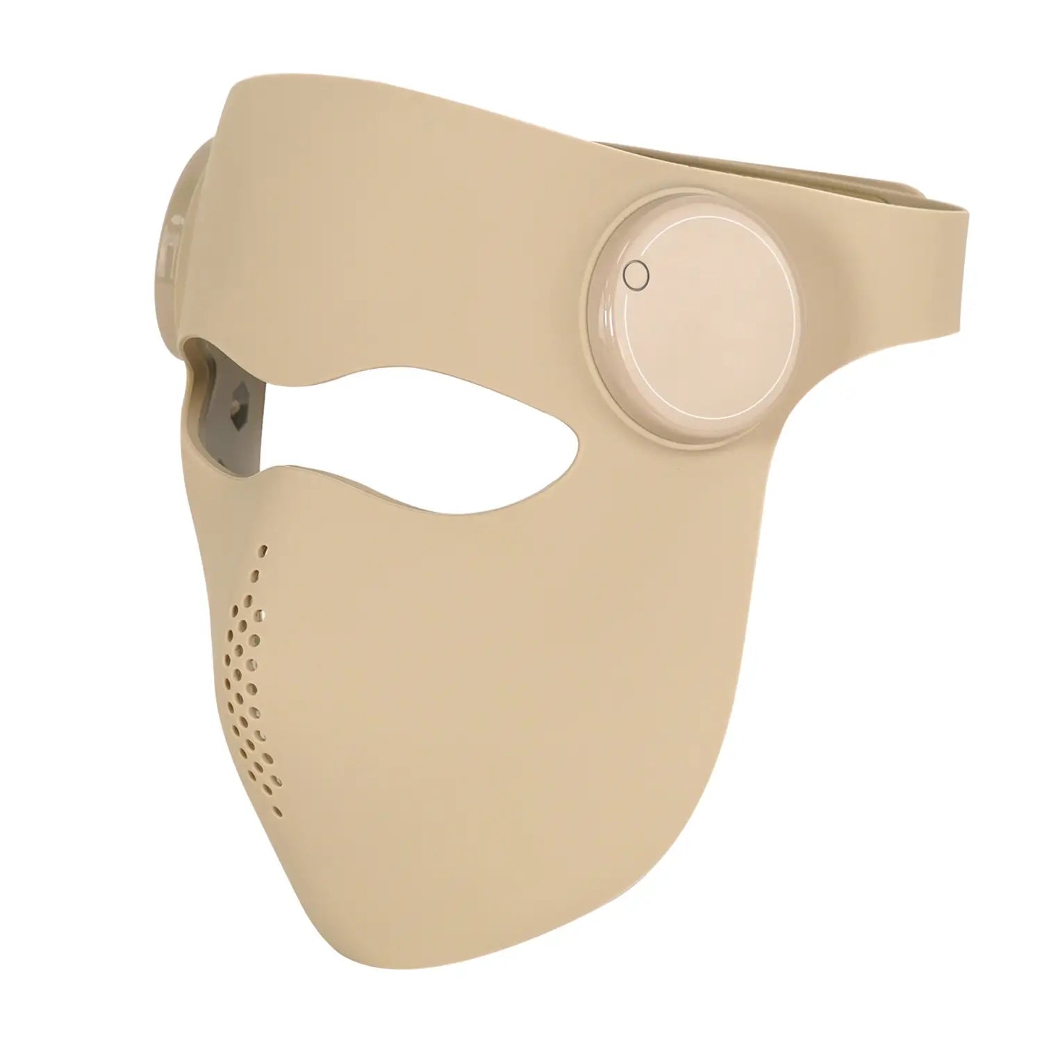
In brain and cognitive health, UCLA and other academic centers have highlighted early studies where intranasal and transcranial near‑infrared light in people with mild to moderate dementia improved cognitive scores over several weeks of treatment and did so without significant adverse effects. The mechanism proposed is exactly the same: red and near‑infrared light penetrate skull bone, are absorbed in brain mitochondria, boost membrane potential and ATP, and reduce local inflammation.
All of this is upstream of the plasma membrane potential that textbooks talk about, but you cannot separate them. The sodium‑potassium pumps that maintain your neuron’s resting potential run on ATP. If mitochondrial membrane potential is depressed, ATP production falls, and the cell struggles to keep its outer membrane polarized. Supporting mitochondrial voltage is therefore one of the cleanest ways to indirectly support overall cell membrane potential.

Practical Ways To Use Red Light To Support Membrane Potential
From an optimization perspective, the question is how to turn all this into a protocol you can actually use without either under‑dosing or frying your mitochondria with too much ROS. The common thread across reputable sources, from Cleveland Clinic to US military photobiomodulation research and the immunomodulation review, is to treat light like a drug with dose, frequency, and target all carefully defined.
Wavelength comes first. Most of the mitochondrial and membrane potential data sit between about 600 and 900 nanometers. Shorter red wavelengths around 633 to 670 nanometers tend to be best for skin, superficial connective tissue, and some immune applications. Deeper near‑infrared wavelengths around 808 to 850 nanometers penetrate further, reaching muscle, joints, and in some studies even spinal cord and brain tissue. A USU pain research team has identified around 810 nanometers as an especially effective wavelength for deep penetration and nerve modulation, calling that range a therapeutic window.
Dose and irradiance come next. The immunomodulation review reports effective fluences around 4 to 12 joules per square centimeter in autoimmune and inflammatory trials. Rheumatoid arthritis patients saw strong benefits at 10 joules per square centimeter three times per week. Chronic wounds responded to 4 joules per square centimeter daily. Asthma improvements were seen at 6 joules per square centimeter twice weekly using a 904 nanometer source. A fibroblast study showed that 5 to 10 joules per square centimeter at 636 or 825 nanometers produced significant metabolic shifts without killing cells, while higher doses pushed ROS much higher.
To translate this into a home routine, you need to know the irradiance of your device at the distance you actually use. If a panel delivers about 10 milliwatts per square centimeter at the distance where your skin sits, a ten‑minute session gives you about 6 joules per square centimeter, right in the low‑to‑moderate window of many successful trials. If your device is stronger, you can reduce time accordingly.
The most evidence‑based applications for now are skin rejuvenation, hair thinning in non‑bald areas, early wound healing, specific joint or tendon pain, and carefully supervised ophthalmic and neurological uses. Stanford dermatology experts emphasize that red light can modestly improve wrinkles and skin thinning and help hair regrowth in areas where follicles are still alive, but does not rebuild dead follicles and is not a magic anti‑aging cure. Cleveland Clinic stresses that evidence for weight loss, cellulite, and broad mental health claims remains weak or nonexistent.
Consistency matters more than hero doses. The immunomodulation and sports recovery data show that benefits usually emerge over six to twelve weeks of repeated use rather than after single marathons. UCLA’s dementia work found cognitive improvements with six minutes per day over eight weeks, not with occasional long sessions. Mitohealth’s myopia and retinal studies use multiple brief exposures per week. In practice, shorter, frequent sessions aligned with your circadian rhythm, such as morning or late‑afternoon exposures, tend to be kinder to membranes than rare, intense blasts.
Eye and skin safety should never be an afterthought. Cleveland Clinic and the American Academy of Dermatology advise protecting your eyes around bright red and near‑infrared sources unless you are under ophthalmic supervision, and they warn that darker skin tones may be more prone to hyperpigmentation if overtreated. The fibroblast and keratinocyte literature also reminds us that beyond a certain dose window, ROS can outstrip antioxidant defenses and damage membranes rather than support them.
A simple strategy that aligns with the published data is to start at the low end of time and frequency, pay attention to how your sleep, energy, and recovery respond over a few weeks, and only then decide whether to step up exposure.
Here is a high‑level comparison of wavelength bands and their most studied effects on mitochondrial and membrane function, synthesized from the immunomodulation review, fibroblast and connective tissue studies, and ophthalmic work.
Wavelength band |
Typical targets |
Evidence themes for membrane potential and mitochondria |
630–670 nm |
Skin, superficial connective tissue, retina |
Increases ATP and mitochondrial membrane potential in fibroblasts and retinal cells; supports collagen production and early wound healing; improves vision metrics in older adults with very brief exposures. |
780–850 nm |
Deeper muscle, joints, spine, brain |
Strong penetration; boosts ATP and modulates pain signaling; improves outcomes in rheumatoid arthritis, inflammatory bowel disease, and asthma at carefully dosed fluences; enhances nerve function in preclinical spinal cord models. |
Around 904 nm |
Respiratory tissue and joints |
Used in asthma and some musculoskeletal trials with reductions in exacerbations and pain; mechanism similar but penetration characteristics differ. |
These are not rigid prescriptions, but they illustrate how different wavelengths interact with different depths and tissues and why “just buy any red light” is not a serious optimization strategy.
Pros, Cons, And Who Should Be Cautious
On the plus side, red light therapy aligns almost perfectly with a systems‑level, non‑pharmaceutical wellness model. It is non‑invasive, generally comfortable, and, when dosed correctly, has a strong safety profile. Cleveland Clinic notes that red and near‑infrared light are non‑ionizing and do not carry the DNA damage risk of ultraviolet light. The immunomodulation review found no significant adverse effects even in multi‑week autoimmune and inflammatory trials with thousands of sessions combined. US military pain research has shown that properly dosed near‑infrared therapy can match the analgesic effects of peripheral nerve injections without impairing motor function.
From a membrane‑centric viewpoint, red light therapy is one of the few tools that directly supports the machinery that maintains cell and mitochondrial membrane potential without drugs or extreme interventions. It sits alongside sleep, nutrition, and physical activity as a foundational input rather than a band‑aid.
The downsides are equally important for an honest, evidence‑based view. The Stanford dermatology piece points out that claims around athletic performance, muscle recovery, and sleep are still speculative, with current data weak or inconsistent despite strong theoretical rationale. Cleveland Clinic emphasizes that many online promises around weight loss, cellulite reduction, and mental health cures do not have good scientific backing.
Device quality and parameter reporting are another weak link. Consumer panels vary widely in wavelength accuracy, irradiance, and build quality. Multiple reviews in photomedicine journals have called out poor reporting of key parameters and wide variability between devices, making it hard to translate clinical doses to home units.
Finally, there are people who should proceed only under medical guidance. Anyone with a history of photosensitive conditions or who is taking photosensitizing medications should be screened before treatment, as recommended in regenerative medicine and safety reviews. Individuals with darker skin tones may need extra caution around facial devices to avoid hyperpigmentation, as dermatology societies have noted. Ophthalmic applications for myopia or retinal disease should always use purpose‑built devices under professional supervision.
How To Think About Red Light As A Membrane‑Level Biohack
If you strip away the marketing and keep only what is durable across Cleveland Clinic, Nature, PubMed, and the better clinical trials, a simple picture emerges. Red and near‑infrared light in very specific windows can increase mitochondrial membrane potential, ATP production, and redox balance in a way that nudges cells back toward a high‑readiness, low‑inflammation state. That shift in mitochondrial voltage cascades into healthier cell membrane potential, cleaner signaling, and better repair.
This does not make red light therapy a cure‑all. It does make it one of the more elegant tools for supporting the electrical and energetic foundation beneath your training, nutrition, and sleep protocols. If you approach it like a veteran optimizer would approach a new supplement—respect the dose window, start low, track real‑world metrics, and stay grounded in what the literature actually shows—you can use light to quietly reshape the membrane potential story inside your cells.

References
- https://digitalcommons.kansascity.edu/cgi/viewcontent.cgi?article=2015&context=studentpub
- https://mavmatrix.uta.edu/cgi/viewcontent.cgi?article=1212&context=bioengineering_theses
- https://ui.adsabs.harvard.edu/link_gateway/2022Photo...9..618S/PUB_HTML
- https://pmc.ncbi.nlm.nih.gov/articles/PMC11991943/
- https://news.usuhs.edu/2022/10/usu-professor-researches-novel-pain.html
- https://admisiones.unicah.edu/uploaded-files/AQgyUl/2OK044/mitochondria__red_light-therapy.pdf
- http://web.stanford.edu/group/barronlab/PubPdfs/2023/The%20anti-inflammatory%20effects%20of%20photobiomodulation%20are%20mediated%20by%20cytoki...ation..pdf
- https://deepblue.lib.umich.edu/bitstream/handle/2027.42/167025/iub2405_am.pdf?sequence=1
- https://my.clevelandclinic.org/health/articles/22114-red-light-therapy
- https://www.uclahealth.org/news/article/5-health-benefits-red-light-therapy