
When you spend as much time around light therapy as I do, you end up seeing it in every corner of recovery: joint pain, brain fog, hair regrowth, stubborn scars. But burns and scalds are in a different league. They are painful, high-stakes injuries where days or even hours matter for infection risk, healing time, and scarring.
So let’s tackle the question that every light-therapy geek eventually asks after a kitchen mishap or a hot water accident: can red light therapy realistically play a role in the emergency treatment of burns and scalds — and if so, how, and when, without doing something reckless?
I’ll walk you through what the lab and clinical data actually say, where the evidence is strongest, and how I personally integrate red light therapy as a second‑line tool after proper burn first aid and medical assessment, not instead of them.
What Red Light Therapy Really Does in Burned Tissue
Red light therapy, also called photobiomodulation or low-level light therapy, uses visible red light (roughly 620–700 nanometers) and near‑infrared light (roughly 800–1000 nanometers) at non‑heating intensities. These wavelengths can penetrate skin and are absorbed by several key targets inside your cells.
From decades of work that began with NASA and has continued through hundreds of studies summarized by groups such as the Cleveland Clinic, MD Anderson Cancer Center, Stanford dermatology, and multiple wound‑healing reviews, a consistent mechanistic picture has emerged.
Mitochondria, Cytochrome c Oxidase, and ATP
The primary photoreceptor in this range appears to be cytochrome c oxidase, a mitochondrial enzyme. When red or near‑infrared photons hit it, several things tend to happen:
Mitochondria make more ATP, the cell’s energy currency. Red light also modulates the redox state of the respiratory chain and releases bound nitric oxide, which improves microcirculation. Studies covered in regenerative biology reviews report increases in oxygen consumption, RNA and protein synthesis, and membrane potential after dosing with low‑level red light.
If you have a burn, the cells you care most about — fibroblasts, keratinocytes, endothelial cells, immune cells — are all energy‑hungry. More ATP and better microcirculation give them more capacity to repair tissue instead of staying stuck in damage control.
Fibroblasts, Collagen, and Angiogenesis
Burn healing depends heavily on fibroblasts. They migrate into the wound, proliferate, and lay down collagen to form granulation tissue. Several converging lines of evidence show that red light has direct, dose‑dependent effects on those cells.
An in‑vitro fibroblast scratch model using 661‑nanometer laser light tested a range of doses from about 1.5 to 11.7 joules per square centimeter. All tested doses were non‑toxic, but the fastest wound closure at 24 hours occurred in a fairly narrow band around 3 to 4.5 joules per square centimeter, with significantly increased fibroblast migration and proliferation compared with unlit controls.
Other work with 660‑nanometer LEDs on normal and diabetic skin fibroblasts found that diabetic fibroblasts, which normally struggle to migrate and produce collagen, actually improved their survival, movement, and collagen production after exposure. That is crucial because diabetic burns and ulcers are notoriously slow to heal.
At the tissue level, multiple studies have reported increased collagen type I production and higher tensile strength in wounds treated with red or near‑infrared light. For example, a review of controlled animal wound models across a range of wavelengths concluded that light therapy significantly improved wound contraction and tensile strength. A separate visible‑light study at 630 nanometers showed upregulation of COL1A1 and COL2A1 (collagen genes) and VEGF (a key angiogenesis factor), while lowering the inflammatory cytokine IL‑1β, in mouse wound models over about three weeks.
Taken together, these data explain why burn and surgical wounds in red‑light‑treated animals often close faster and with stronger, better‑organized tissue.
Inflammation, Growth Factors, and Signaling Pathways
Burns are not just mechanical injuries; they are inflammatory storms. Photobiomodulation consistently shifts that storm toward a more constructive pattern.
Experimental and clinical work shows that red and near‑infrared light can reduce pro‑inflammatory cytokines and increase anti‑inflammatory cytokines. A University at Buffalo–led mouse study on third‑degree burns demonstrated that low‑dose photobiomodulation accelerated healing and reduced inflammation by activating TGF‑beta 1, a growth‑regulating protein that orchestrates both immune responses and tissue repair. That activation in turn stimulated fibroblasts and macrophages, the cells that build tissue and clean up debris.
Other research has tied visible and near‑infrared light to modulation of STAT3 signaling, a key transcription factor for keratinocyte proliferation, angiogenesis, and fibrosis. When properly tuned, these pathways support faster closure and less scarring; when misregulated, they can drive pathological scarring or unchecked growth. This is why dose and timing matter so much.
The Goldilocks Dose Problem
Across wound and burn models, red light follows a biphasic dose‑response curve, often described as the Arndt–Schulz law. Very low doses do almost nothing. Moderate doses stimulate repair. Higher doses gradually lose benefit and can even inhibit healing.
You see this clearly in the 661‑nanometer fibroblast model: everything benefited relative to control, but the sweet spot for accelerated wound closure sat around 3 to 4.5 joules per square centimeter. In burn‑focused reviews and protocols, energy densities in the single‑digit to a few tens of joules per square centimeter range are often effective, while doses above roughly 50 joules per square centimeter become less reliable or potentially counterproductive.
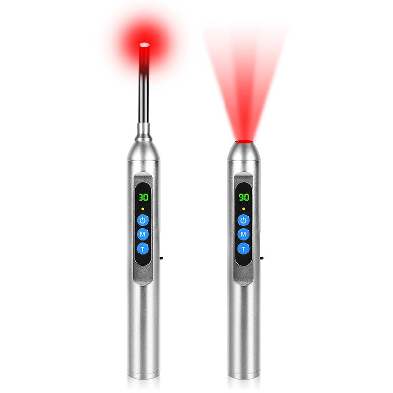
Consumer‑facing guides, like the educational materials from Atria and several health systems, translate this into practical parameters: devices that deliver about 20 to 100 milliwatts per square centimeter at the skin, for roughly 5 to 20 minutes per area, at distances around 6 to 24 inches, depending on the power of the panel.
A quick example demonstrates why this matters. If a small panel delivers about 40 milliwatts per square centimeter at 12 inches, then every second provides 0.04 joules per square centimeter. To hit roughly 4 joules per square centimeter — the zone that helped fibroblast closure in that lab model — you would need about 100 seconds, just over 1.5 minutes. If you stood there for 20 minutes at the same distance, you would deliver about 48 joules per square centimeter, far into the “more is not better” territory many wound papers warn about.
This is the core of safe and effective light use for burns: enough energy to signal regeneration and calm inflammation, not so much that you blunt the response.

What the Evidence Says Specifically About Burns and Scalds
Now, the question you really care about: what do real burn and wound studies show when red light is added?
Animal and Lab Work: Proof of Principle
Animal studies are not the finish line, but they are the proving ground for mechanism and dose.
In third‑degree mouse burns, the University at Buffalo protocol applied carefully titrated photobiomodulation and saw faster closure over nine days and less inflammation, driven by TGF‑beta 1 activation. This is deep, full‑thickness injury, not just a kitchen splash.
Burn‑wound systematic reviews looking at about two decades of experimental work have reached similar conclusions. One review of 22 burn studies reported that low‑level laser or LED therapies accelerated healing across different animal models, improving new tissue formation and reducing inflammatory damage. Another analysis reported that second‑degree burns responded best when red light was introduced during the proliferative phase, the rebuilding stage after the immediate crisis of hemostasis and acute inflammation.
In vitro, the fibroblast scratch models at 661 nanometers showed that the right dose of red light activates both migration and proliferation without toxicity. At the molecular level, red light increased ROS in a controlled way, nudging the signaling pathways that drive cell movement into the wound gap. These are the same cells you depend on to rebuild burned dermis.
All of this explains why both NASA‑initiated wound work and later commercial systems have leaned hard into red and near‑infrared bands for tissue repair.
Human Burn and Postsurgical Data: Small but Encouraging
Direct human burn research is more limited, but it exists and is growing.
One early LED study using 658‑nanometer red light in five people with bilateral burns treated one side and used the contralateral side as a control. The irradiated side re‑epithelialized faster than the unexposed side, suggesting a local effect beyond dressing care alone.
A more recent randomized controlled trial protocol out of Brazil, registered in a national trial registry, is currently testing three arms in adults with second‑degree burns: red LED at 658 nanometers, infrared LED at 858 nanometers, and a sham treatment delivering minimal energy. Thirty‑nine participants are being followed until complete re‑epithelialization, with daily measures of wound size, pain, itching, skin temperature, and later scar quality and mobility. Before this protocol, low‑level laser burn studies in humans were rare; this trial is designed specifically to lock down which wavelengths and doses actually matter for real‑world burns.
Case‑level data add some color. A Korean case report described a patient with diabetes and a second‑degree burn treated with 830‑nanometer LED phototherapy combined with topical epidermal growth factor. Despite diabetes usually delaying healing, the wound fully re‑epithelialized in ten days, with only mild redness and pigmentation afterwards. Clinically, most second‑degree burns heal in about two weeks when everything goes well, so shaving several days off in a high‑risk patient is meaningful.
On the severe side, a 2016 case series in diabetic patients with third‑degree burns combined red and near‑infrared photobiomodulation with split‑thickness skin grafting. These were individuals initially considered for amputation. After about eight weeks, all participants healed their grafts without amputation. That is not a randomized trial, but it shows that even deep, compromised burns can respond when light is layered onto sound surgical management.
Beyond burns, larger wound populations give us more confidence in the general wound‑healing effect. In one double‑blind study of chronic leg ulcers in seventy‑nine patients, those receiving LED therapy had substantially better increases in blood microcirculation and healing compared with controls. Diabetic leg‑ulcer studies and reviews in journals such as Lasers in Surgery and Medicine and Regenerative Biology report similar themes: improved closure rates, smaller ulcer size, better granulation tissue, and better pain control.
Post‑surgical incisions tell the same story from a different angle. In a trial of ninety sternotomy patients, those receiving red‑LED therapy experienced less pain, fewer wound complications, and easier recovery than controls. In another study on dogs after back surgery, laser‑treated wounds looked better by day seven and significantly improved by day twenty‑one compared with untreated incisions.
Scars and Burn Sequelae
Emergency care is about survival and immediate closure; long‑term quality of life depends heavily on scars.
Several burn‑scar and hypertrophic‑scar studies have used red light after the acute phase. In one pediatric study, each child’s hypertrophic scar was split, with half treated with red light for three months and the other half left untreated. The treated half showed noticeably less raised, less visible scarring. A 2004 burn‑scar study reported roughly twice the decrease in visible scarring in red‑light‑treated scars compared with no therapy, with no reported adverse effects.
A case report of a fifteen‑centimeter cholecystectomy scar treated with photobiomodulation documented improvements in both appearance and neuropathic pain, reinforcing the idea that light can help not just how a scar looks but how it feels.
Taken together, small human trials, case reports, and animal models align: when the dose is right and the therapy is started at an appropriate phase, red‑spectrum light can accelerate burn‑wound closure, support better collagen architecture, improve microcirculation, and likely reduce long‑term scarring.
Where Red Light Fits in Emergency Burn Care
This is where we need to be brutally clear. Red light therapy is not the first thing you reach for when your skin is still hot, blistering rapidly, or charred. The first moves in any burn are still basic emergency care and triage, then evidence‑based wound management. Red light belongs as an adjunct, not a replacement.
Step Zero: Standard Burn First Aid and Medical Assessment
For any significant burn or scald, the first priorities are to stop the burning process, gently cool the area, and seek medical evaluation. That includes removing the heat source, getting away from steam or hot fluids, removing tight jewelry or clothing near the area if possible, and letting a professional decide whether you are dealing with a minor superficial injury or something that needs a burn unit.
No light therapy device, no matter how expensive or science‑y, can debride dead tissue, deal with smoke inhalation, reverse shock, or substitute for tetanus shots, antibiotics, or surgery. The best clinical burn trials that used red or near‑infrared light all layered it on top of standard care, not instead of it.
When Red Light Can Enter the Picture
Where red light does start to make sense in an emergency‑to‑recovery timeline is after three conditions are met.
First, the burn has been cooled and medically assessed. Second, the skin is no longer actively being destroyed by heat or chemicals. Third, the plan for dressings, possible grafts, and infection prevention is in place.
For minor, superficial burns and scalds, think about the small, intensely painful kitchen burns many of us have had, adding a properly dosed red‑light panel or handheld LED in the first day or two can be a reasonable way to try to reduce pain and support faster healing. This is consistent with broader clinical experience that low‑level light can reduce acute pain and calm inflammation in soft‑tissue injuries.
For deeper partial‑thickness, blistering burns and any full‑thickness or circumferential injuries, red light belongs in a supervised setting, if at all. That might mean a wound clinic, a burn center, or a physical therapy department that already uses LED photobiomodulation as part of its protocol.
In those settings, clinicians can choose wavelengths and energy doses similar to the ones studied in burns and chronic wounds, monitor daily re‑epithelialization, track pain and itching, and adjust treatment based on observable changes.
At‑Home Panels vs Clinical Devices
One of the most important practical questions is whether you can just grab the same panel you use for skin rejuvenation or joint pain and point it at a fresh burn.
Here is the core reality that organizations like the Cleveland Clinic, WebMD, and MD Anderson all emphasize: at‑home devices are usually far less powerful and rarely validated for wound or burn indications. Some are cleared by the FDA for issues like facial wrinkles or hair loss, but that clearance primarily addresses safety in that use, not confirmed efficacy for acute burns.
By contrast, clinical devices used in trials and wound centers are designed around specific parameters. Examples include:
An Endophoton system set at 658 nanometers with defined power per diode and an energy density of about 3 joules per point, spaced roughly 0.8 inches apart, delivered until full re‑epithelialization in second‑degree burns.
A combination device using 660‑nanometer LEDs and 808‑nanometer infrared lasers to deliver both superficial and deeper tissue energy in postsurgical and joint contexts.
An 830‑nanometer LED system paired with epidermal growth factor solution in a diabetic burn patient.
A single device like the Dior × Lucibel 630‑nanometer LED mask that delivers about 15.6 joules per square centimeter in a carefully timed twelve‑minute session, validated for facial photoaging rather than burns but illustrating how tightly some manufacturers control dose.
The bottom line is that well‑run burn and wound studies use medical‑grade light systems with known wavelengths, irradiance, and fluence, often under daily or near‑daily supervision. Most consumer panels and masks simply are not engineered or cleared to deliver that kind of precise wound‑healing protocol.
A practical way to think about it is to treat at‑home red light on a minor burn as a low‑risk experiment at conservative doses and to insist on professional devices and guidance for anything beyond that.
A simple comparison helps crystallize it:
Scenario |
Role for Red Light |
Evidence Level |
Best Action |
Small superficial kitchen burn on healthy adult |
Adjunct for pain relief and faster healing after basic first aid |
Indirect, extrapolated from general wound data |
Consider short, moderate‑dose panel or LED use if skin is intact, while monitoring closely |
Deeper partial‑thickness burn, large scald, or any burn in diabetes or vascular disease |
Potential adjunct to improve healing and reduce complications |
Case reports, small series, emerging RCTs |
Use only within a wound clinic or burn center that already employs LED/laser protocols |
Full‑thickness burn, electrical or chemical burn, or signs of systemic illness |
Red light not first‑line; may have a role later in rehab or scar management |
Very limited, mostly animal and case‑level |
Prioritize emergency and surgical care; discuss light therapy later with the burn team |
Practical Protocol Thinking for Light‑Savvy Burn Recovery
If you are already living the biohacking lifestyle and want to integrate red light intelligently once the initial crisis passes, here is how to align with the science without overreaching.
Wavelength Strategy
Most burn and wound‑healing work clusters in three wavelength bands.
First, mid‑red, roughly 630 to 670 nanometers. NASA‑linked wound work, diabetic fibroblast studies, and chronic‑wound clinical trials frequently use 660 to 670 nanometers. In an in‑vitro comparison of several low‑intensity lasers, fibroblasts multiplied fastest with light at about 665 to 675 nanometers, while 810 nanometers actually slowed their proliferation.
Second, near‑infrared around 800 to 860 nanometers. Devices using 808 or 858 nanometers are common in joint and deep‑tissue protocols. In burn contexts, near‑infrared appears to help deeper structures, improving circulation and collagen in full‑thickness or grafted wounds.
Third, combined systems. Some devices deliberately pair 660‑nanometer LEDs with 808‑nanometer lasers to address both surface and deeper tissues. Case reports and product‑linked series suggest that this combination can reduce pain and support faster function recovery after surgery and injuries.
For superficial and partial‑thickness burns, evidence leans more heavily on red rather than pure near‑infrared light. For deep burns, combined protocols with near‑infrared may be rational, but those should be designed and supervised by clinicians.
Dose and Duration: Thinking in Joules, Not Minutes
The studies that give us actual numbers talk in joules per square centimeter, not “fifteen minutes on level three.”
Lab and protocol data suggest that for integumentary healing, doses in the 3 to 6 joules per square centimeter range, and in some chronic wounds up to a few tens of joules per square centimeter, can be effective. Above about 50 joules per square centimeter, benefits tend to plateau or reverse.
To translate that into real‑world sessions, you need at least an approximate power density. Many mid‑range panels deliver roughly 20 to 100 milliwatts per square centimeter at a distance of 6 to 24 inches, as described in practical guides from light‑therapy educators.
Using those numbers, if your device delivers about 50 milliwatts per square centimeter at 8 inches, then every second delivers 0.05 joules per square centimeter. Hitting 5 joules per square centimeter would take about 100 seconds; hitting 25 joules per square centimeter would take roughly 8 minutes and 20 seconds.
You can see why the fibroblast model’s 3 to 4.5 joule per square centimeter sweet spot might translate to a couple of minutes at moderate intensity rather than twenty minutes pressed right against the skin.
For actual burn treatment, especially in a clinic, the protocol will also adjust for wound size, position, dressing type, and how the tissue is responding over days. That is not something you can fully recreate at home, but thinking in terms of energy dose instead of only time and brightness will keep you closer to the therapeutic window.
Frequency and Timing Across the Healing Timeline
Healing is not linear, and neither is optimal light dosing.
In the Dior × Lucibel facial antiaging study, twice‑weekly twelve‑minute sessions over three months produced measurable improvements in wrinkles, firmness, and dermal density, with benefits persisting for about a month after stopping. Chronic‑wound and leg‑ulcer trials often use near‑daily sessions in the early weeks, then taper as closure progresses.
In burns, experimental third‑degree mouse work used daily exposures across a nine‑day healing window. The diabetic third‑degree human series combined photobiomodulation with standard graft protocols over about eight weeks. The pediatric scar trial treated half‑scars for three months.
In practical terms, that suggests more frequent but moderate dosing in the early days and weeks of burn healing, followed by less frequent, lower‑dose work when you are primarily concerned with scar remodeling rather than raw closure. This mirrors how wound clinics schedule debridement and dressing changes: more intense early, then less as the tissue stabilizes.
Pros, Cons, and Unknowns for Emergency Burn Use
When you step back from the enthusiasm and look at red light for burns the way a conservative dermatologist or burn surgeon would, the picture is nuanced.
On the plus side, we have:
Non‑invasive, non‑ionizing, non‑UV light that does not add thermal injury at the doses used in wound healing, which is why NASA, cancer‑care organizations, and major medical centers use it in contexts as sensitive as oral mucositis and retinal disease support.
Hundreds of trials in wounds, ulcers, pain conditions, and skin aging reporting improved tissue repair, reduced pain, better collagen architecture, and reduced inflammation, with wound‑specific reviews concluding that light therapy can increase wound tensile strength and contraction and speed closure.
Burn‑specific animal and early human data showing faster re‑epithelialization, better collagen types I and III deposition, reduced inflammatory damage, and even limb‑saving outcomes in challenging diabetic cases.
A strong safety profile in the short term when devices are used properly, with side effects usually limited to mild, transient redness or warmth. Major organizations explicitly note that red light does not behave like ultraviolet and is not documented to cause skin cancer.
On the minus side, we have:
Heterogeneous protocols and small sample sizes. Many burn and wound studies are underpowered, single‑center, or case‑based. The ongoing randomized trials in second‑degree burns are promising because they are designed to tighten this up, but results are still emerging.
Lack of standardized dosing. Even within the same wavelength, different studies use different irradiances, exposure times, and session frequencies. The biphasic response means that simply “adding more minutes” is not a safe heuristic.
Unclear best practices for the true emergency window. Almost all evidence introduces light after basic burn stabilization and wound‑care decisions are made. We do not have solid data for pointing a consumer panel at a fresh blister within minutes of injury. Caution is warranted here.
Device variability. Clinical LED and laser systems document their exact power density and beam profile at the treatment distance. Home devices rarely provide accurate irradiance maps, and some deliver very low power that may never reach therapeutic fluences without unreasonably long sessions.
Psychological risk. Perhaps the biggest danger for biohackers is overconfidence. If someone tries to “treat” a serious burn at home with light therapy instead of going to a burn unit, no amount of mitochondrial activation will make up for missed antibiotics, surgery, or proper dressings.
The responsible position — and it is the one taken by mainstream sources like the Cleveland Clinic, MD Anderson, Stanford dermatology, and WebMD — is to frame red light as a promising adjunct with decent safety and mechanistic plausibility, backed by encouraging but not definitive evidence for wound and burn healing. It is not a stand‑alone emergency solution.

How I Actually Use Red Light Around Burns
Speaking as a veteran wellness optimizer, here is how I integrate all this when a burn shows up in real life.
For small, superficial burns and scalds that have already been cooled and assessed as minor, I treat red light as a second‑line tool for comfort and optimization. I will use a mid‑red device with known wavelengths in the mid‑600s, at a comfortable distance where the skin feels warm at most, not hot, for a few minutes at a time, once or twice a day in the first few days. The goal is to fall somewhere near the low single‑digit to low double‑digit joules per square centimeter zone, not to blast the area. I pay more attention to how the tissue looks and feels over twenty‑four to forty‑eight hours than to the exact minute count.
For anything deeper, larger, or in someone with diabetes, vascular disease, or immune compromise, my threshold for involving a wound clinic or burn specialist is very low. If that team already uses LED or laser photobiomodulation, I am happy; if they do not, I will share the relevant literature and let them decide whether and how to integrate it. In this range of burns, I do not self‑prescribe light but I do strongly advocate for its inclusion as part of a research‑backed, multidisciplinary regimen.
For scars and chronic post‑burn pain, red light becomes a long‑term remodeling partner rather than an emergency tool. The hypertrophic scar data, neuropathic‑scar case reports, and anti‑aging facial studies all suggest that gently nudging collagen and microcirculation week after week, for months, can shift scar texture, color, and sensation in the right direction.
In every scenario, the meta‑rule is the same: treat light like a powerful, subtle signal to biology. Use it to support what your body and your burn‑care team are already trying to do — not as a shortcut around fundamentals like cooling, cleaning, debridement, dressings, grafts, and infection control.
If you respect those boundaries, red light therapy can move from a wellness gadget into a genuinely useful tool for emergency‑to‑recovery burn care, especially for those of us who are willing to think in wavelengths, joules, and healing timelines rather than just in minutes under a glow.
References
- https://pubmed.ncbi.nlm.nih.gov/40175683/
- https://www.buffalo.edu/news/releases/2021/08/003.html
- https://med.stanford.edu/news/insights/2025/02/red-light-therapy-skin-hair-medical-clinics.html
- https://www.brownhealth.org/be-well/red-light-therapy-benefits-safety-and-things-know
- https://www.mdanderson.org/cancerwise/what-is-red-light-therapy.h00-159701490.html
- https://atria.org/education/your-guide-to-red-light-therapy/
- https://my.clevelandclinic.org/health/articles/22114-red-light-therapy
- https://www.gundersenhealth.org/health-wellness/aging-well/exploring-the-benefits-of-red-light-therapy
- https://www.uclahealth.org/news/article/5-health-benefits-red-light-therapy
- https://www.aad.org/public/cosmetic/safety/red-light-therapy