
As someone who has spent decades biohacking recovery protocols for athletes, post-op patients, and my own stubborn scars, I can tell you this: red light is not magic, but it behaves uncannily like a well-designed drug. It has a dose, a target, a mechanism, and side effects if you push it too far. When you respect that, you can tilt wound healing away from thick, raised scar tissue and toward quiet, almost invisible repair.
This article walks through how red light actually changes wound biology, what the strongest studies say about scarring, and how to think about wavelength, dose, and timing if you want real, science-backed results rather than Instagram placebo.
Photobiomodulation 101: How Red Light Talks To Your Cells
Red and near‑infrared light therapy is often called photobiomodulation or low‑level light therapy. Cleveland Clinic describes it as low‑power red or near‑infrared light that works not by heating tissue, but by being absorbed in mitochondria, boosting cellular energy, modulating inflammation, and stimulating collagen production. Harvard Health echoes this, noting that the leading theory is mitochondrial stimulation that leads to reduced inflammation and stronger, more supple skin.
One of the most important mechanistic papers came out in the journal Mitochondrion in 2004. The authors showed that red and near‑infrared light in roughly the 630–1,000 nm range interacts directly with cytochrome c oxidase, a key mitochondrial enzyme. When this enzyme absorbs photons, several things happen: oxidative metabolism is enhanced, ATP production rises, and gene expression pathways involved in antioxidant protection and energy production are upregulated. In diabetic mice, this same kind of light upregulated genes tied to tissue repair and measurably improved wound healing.
From an engineer’s point of view, you can think of this as an input (photons) that nudges a metabolic control system. The first‑order effect is more ATP. The second‑order effects show up as more collagen synthesis, faster cell migration, and better antioxidant defenses. A review of 68 preclinical LASER and LED wound studies found consistent patterns: red and near‑infrared light reduced inflammatory cells, increased fibroblast and keratinocyte proliferation, stimulated angiogenesis, and enhanced granulation tissue formation. Those are exactly the levers you care about if your goal is fast healing with minimal scarring.
To make this concrete, consider a well‑controlled fibroblast study that used a 661 nm diode laser on mouse 3T3 cells. At a power density of 10 mW/cm² for 8 minutes, the cells received about 4.8 J/cm² of energy. That single, short exposure significantly accelerated “wound” closure in a scratch assay at 24 hours, compared with non‑irradiated controls, without any phototoxicity. In parallel, reactive oxygen species (ROS) and cell orientation toward the wound margin increased in a way that lined up with faster closure. That is not mystical; it is a dose of light creating a quantifiable change in cell behavior.

Wound Healing, Step By Step – And Where Red Light Fits
Every skin wound follows the same four overlapping phases: hemostasis, inflammation, proliferation, and remodeling. Red light does not replace that choreography; it tunes it. The most exciting work on scar reduction shows that red and visible light can accelerate early repair while dialing down the signals that drive chronic inflammation and excessive fibrosis.
Inflammation: Reducing The “Volume” Without Muting The Signal
Early inflammation is essential. White blood cells clean up debris and microbes, and cytokines coordinate the response. The problem is not the initial burst; it is when inflammatory signaling stays loud for too long. That is how you get chronic, stalled wounds and thick scars.
A recent mouse study of 630 nm red light focused on this stage. After optimizing parameters in vitro using NIH/3T3 fibroblasts, the researchers created standardized full‑thickness wounds about 4 mm across (roughly 0.16 in) on nude mice and treated them with red light. By three weeks, red‑light‑treated wounds showed significantly higher expression of COL1A1 and COL2A1 (genes encoding type I and II collagen) and vascular endothelial growth factor (VEGF), along with lower levels of the pro‑inflammatory cytokine IL‑1β. In plain language, the wounds had more organized collagen, better angiogenesis, and quieter inflammation compared with controls, with effects comparable to a hydrogel positive control.
The big picture here matches what a variety of sources report. Lumitex, summarizing photobiomodulation for wound healing, notes that red and near‑infrared light reduce inflammation while improving blood flow and collagen production. DegreeWellness reviews controlled trials in post‑sternotomy patients where red light reduced pain, bleeding, wound ruptures, and blood‑related complications. A Nature Communications paper looking at visible light in mice goes one level deeper, linking visible‑light exposure to modulation of STAT3 signaling, a master transcription factor that integrates cytokine and ROS signals and governs inflammation, angiogenesis, and tissue remodeling.
You do not want zero inflammation. You want a sharp, well‑timed peak and a graceful exit. The animal and molecular data together suggest red light is good at that.
Proliferation: Fibroblasts, Collagen, And Directional Repair
Once the wound is clean, fibroblasts migrate in, proliferate, and lay down new extracellular matrix. Keratinocytes close the surface. This is where red light really shines.
In the 661 nm fibroblast scratch study, fluences in the 3–4.5 J/cm² range produced the fastest closure at 24 hours. The most effective dose, 4.8 J/cm², not only increased viability and migration but also induced fibroblasts to align toward the wound margin, effectively “aiming” their movement and matrix deposition.
Multiple independent lines of evidence converge on this fibroblast story. The preclinical review of 68 LASER and LED experiments found that red/NIR wavelengths in the optical window (mostly 630–830 nm) at low doses around 1–5 J/cm² consistently stimulated fibroblast proliferation and collagen synthesis. In diabetic models summarized by Kineon’s research review, 660 nm light improved motility, survival, and collagen production specifically in diabetic fibroblasts, partially overcoming the impaired healing typical of diabetes.
The 630 nm mouse study mentioned earlier supports this mechanistically: upregulation of COL1A1 and COL2A1 means the matrix being assembled is richer in structural collagens, and VEGF upregulation supports the microvasculature that feeds proliferating cells. Lumitex highlights this same pattern at the tissue level, noting that red and near‑infrared light enhance perfusion, ATP production, and collagen synthesis, which together reduce pain, speed closure, and minimize scarring.
Real‑world examples bring this to life. A 2018 review of controlled wound trials summarized by DegreeWellness found that red light significantly increased tensile strength and wound contraction in animal models, for both open and sutured incisions. In a randomized, double‑blind trial of 90 sternotomy patients, those receiving red light had less pain and fewer wound complications. In a case series of third‑degree burns in diabetic patients treated with skin grafts plus red/near‑infrared light, all patients healed within about 8 weeks with no amputations, despite high baseline risk. That is robust, load‑bearing tissue, not fragile scar that tears on first contact.
Remodeling: Steering The Scar Toward “Invisible”
Remodeling is where the collagen architecture either becomes a thin, flexible, “forgotten” line or a raised, stiff hypertrophic or keloid scar. Here, the evidence is more nuanced but still promising.
On the molecular side, the visible‑light paper in Nature Communications links non‑UV visible light to STAT3‑mediated pathways that govern angiogenesis, macrophage phenotype, and fibroblast behavior. Too much STAT3 activity over the long term is tied to fibrosis and cancer, but tightly controlled activation during acute repair appears essential for proper remodeling. The authors argue that carefully dosed visible light can push STAT3 signaling toward pro‑regenerative modes that improve wound closure and reduce scar formation in mice.
Clinically, DegreeWellness summarizes a 2015 systematic review of 40 plastic‑surgery‑related studies where low‑level laser and red light therapies improved healing of acute wounds and enhanced burn‑scar outcomes in both animal and human work. One pediatric study in 15 children with hypertrophic scars used red light for 3 months on part of each scar and saw significant reductions in thickness and appearance compared with untreated areas. In a 2004 burn‑scar study, red‑light‑treated patients had roughly double the decrease in visible scarring compared with controls, without reported adverse effects.
Stanford dermatology experts reviewing the broader red light literature describe the data on scars as mixed but encouraging. For example, one eyelid surgery study showed faster early scar healing and about 50 percent shorter time for the scar to become minimally visible on treated areas, whereas other small studies found only modest, non‑significant cosmetic differences by six weeks. Harvard Health and the American Academy of Dermatology both acknowledge red light’s potential to speed wound healing and minimize scars, but they stress that protocols are not standardized and results are not guaranteed.
One more fascinating data point comes from aesthetics rather than classic scars. In a clinical trial of a high‑power red LED mask emitting 630 nm light at about 21.7 mW/cm² (dose 15.6 J/cm² over 12 minutes), twice‑weekly sessions for 3 months produced substantial structural changes in facial skin. Dermal density increased by about 47.7 percent, skin roughness dropped roughly 23.8 percent, pore diameter shrank about 32.8 percent, sebum levels decreased around 70 percent, and these improvements persisted for at least a month after stopping treatment. While this study focused on photoaging, not wounds, it shows that properly dosed red light can drive long‑term remodeling of collagen and dermal architecture. That is exactly what you want when you are trying to guide a fresh scar toward “barely there.”

Mechanisms At A Glance
When you put the mechanistic and clinical data side by side, a few core pathways keep showing up.
Mechanism |
Primary targets |
Evidence highlights |
Relevance to scarring |
Mitochondrial activation |
Cytochrome c oxidase, respiratory chain |
Mitochondrion 2004 paper: red/NIR increases mitochondrial activity, ATP, and gene programs for protection |
Higher ATP lets fibroblasts and keratinocytes proliferate and remodel efficiently without energy stress |
Controlled ROS and redox shifts |
ROS signaling, antioxidant defenses |
661 nm fibroblast study: increased ROS at effective doses; multiple PBM reviews note biphasic dose effects |
Moderate ROS acts as a signal for proliferation and repair; too much drives damage and fibrosis |
Collagen gene upregulation |
COL1A1, COL2A1 and related matrix genes |
630 nm mouse study: increased COL1A1, COL2A1 with better histologic repair |
Higher, better‑organized collagen content supports strong, flat tissue rather than weak, raised scars |
Angiogenesis support |
VEGF and downstream vascular pathways |
630 nm mouse wounds: VEGF increased; visible‑light/STAT3 paper links light to VEGF‑STAT3 angiogenesis |
Healthy microvasculature feeds regenerating tissue and reduces ischemic, fibrotic areas |
Inflammation modulation |
IL‑1β, other cytokines, JAK/STAT pathways |
630 nm mouse study: IL‑1β reduced; visible‑light STAT3 modulation; numerous anti‑inflammatory PBM reports |
Shorter, better‑controlled inflammation correlates with fewer hypertrophic and chronic scars |
Fibroblast migration/orientation |
Fibroblast cytoskeleton, ECM interactions |
661 nm scratch assay: faster closure and directional alignment toward wound margin |
Directional repair helps close wounds with minimal distortion and more ordered collagen |
When you understand these pathways, the “scar‑minimizing” effects of red light stop being mysterious and start looking like a natural consequence of well‑tuned cellular control.

Dose, Wavelength, And Timing: Treat Light Like A Drug
The biggest mistake I see biohackers make with red light is assuming that more exposure is always better. The science says the opposite.
A key principle in photobiomodulation is the biphasic dose response, often framed as the Arndt–Schulz law. The review of 68 LASER and LED wound studies found that very low doses did little, doses around 1–5 J/cm² were usually stimulatory, and higher doses around 10–16 J/cm² could actually inhibit cellular functions in some models. The 661 nm fibroblast study supports this: all tested doses increased viability compared with controls, but the highest fluence groups (7.8 and 11.7 J/cm²) did not show the same statistically significant gains as the mid‑range.
A preclinical mouse study using a helium‑neon laser at 632.8 nm adds another nuance. Researchers compared multiple low‑dose exposures of 0.5 J/cm² (delivered up to five times per week) to a single “optimum” 2 J/cm² exposure on full‑thickness wounds about 15 mm across (roughly 0.6 in). Simply stacking more sub‑therapeutic doses did not compensate; multiple 0.5 J/cm² sessions did not improve contraction speed or mean healing time relative to controls, while the single 2 J/cm² dose was clearly superior. In other words, if you are below the effective threshold, doing it more often does not magically fix the dose.
Clinical device designers have come to similar conclusions from another direction. Lumitex reports that many medical protocols for complex wounds use red and near‑infrared fluences in roughly the 5–40 J/cm² range per session, with a caution that doses above about 50 J/cm² can become less effective or even harmful. The Dior × Lucibel mask for facial rejuvenation uses a dose of 15.6 J/cm² per 12‑minute session at an average irradiance of 21.7 mW/cm² and recommends twice‑weekly use with 72 hours between sessions. That protocol produced progressive gains over 3 months without apparent adverse effects, and the authors emphasize the importance of both power density and spacing in respecting the biphasic response.
Wavelength matters just as much as dose. The connective‑tissue review by Shaikh‑Kader notes that red light in the 600–700 nm range and near‑infrared light from about 780–1,100 nm penetrate far more deeply than blue or ultraviolet. Clinical and preclinical wound papers cluster effective wound‑healing wavelengths in the red to near‑infrared window, roughly 630–830 nm. The mitochondrial action spectra reported in the wound‑healing review show peaks near 613–623 nm, 667–684 nm, 751–772 nm, and 813–846 nm, consistent with cytochrome c oxidase absorption.
Anatomy adds another layer. Red light around 630–670 nm is excellent for dermal structures: superficial wounds, surgical incisions, and scars. Near‑infrared around 790–820 nm penetrates deeper; work from Juanita Anders’ group at the Uniformed Services University has shown that 810 nm light can reach the spinal cord transcutaneously and selectively block small pain fibers while changing gene expression in neurons and glia. For deep injuries or joint and bone involvement, near‑infrared becomes more relevant. For many skin wounds and scars, properly dosed red light is sufficient.
Timing is the last parameter people ignore. The Dior mask study explicitly chose twice‑weekly sessions spaced 72 hours apart, arguing that it takes several days for the cellular “energy digestion” and downstream gene expression to play out after a photobiomodulation session. The wound literature backs this idea indirectly: the most effective 661 nm scratch‑assay dose produced its standout results at 24 hours; by 48 hours, all groups, including controls, had nearly closed the scratch, so differences disappeared. Red light seems best viewed as a targeted nudge before the next natural step in the healing sequence, not an all‑day flood.

How All This Translates Into Less Scarring
At this point you might be wondering: does this mechanistic elegance translate into fewer or better scars you can actually see in the mirror? The answer is “often yes, sometimes modestly, rarely dramatically.”
The animal data are strong. The Mitochondrion paper showed that red and near‑infrared light protected retinal and optic‑nerve tissue from toxin‑induced injury and improved wound healing in diabetic mice by upregulating repair genes. Cardiac studies summarized in the Kineon review found that low‑level laser after myocardial infarction reduced mortality in rats and dogs, shrank infarct (scar) size in dogs, and preserved cellular structure and energy stores in rat hearts. Burn models synthesized by DegreeWellness show reduced inflammation, enhanced fibroblast activity, more type I and III collagen, and faster closure in both second‑ and third‑degree burns when red or red/near‑infrared light is applied, especially during the proliferative phase.
Human data are more heterogeneous but increasingly compelling. The sternotomy trial mentioned earlier did not just show better pain scores; it also reported fewer wound ruptures and bleeding episodes, which is a proxy for tissue integrity. The plastic‑surgery systematic review and the pediatric hypertrophic scar trial indicate that repeated red light over months can flatten and soften problematic scars. A small but striking case described in the Kineon review reported a 15 cm (about 6 in) cholecystectomy scar with reduced pain, redness, swelling, and better cosmetic appearance after photobiomodulation.
In chronic wound populations, red and near‑infrared light look particularly valuable. A study of 79 patients with chronic leg wounds found that those receiving LED therapy saw substantial increases in microcirculation and better healing compared with little change in controls. A separate systematic review and meta‑analysis focused on diabetic foot ulcers notes that red and infrared light can improve closure rates as an adjunct to standard care, even though methodological weaknesses and inconsistent parameters have kept these therapies out of formal guideline recommendations so far.
Dermatology perspectives from Stanford, Harvard, Cleveland Clinic, Brown University Health, and others all land in a similar place. Red light can meaningfully modulate biology: boost collagen, smooth texture, improve early scar appearance, and speed closure in some wound types. But protocols differ dramatically in wavelength, power, duration, and frequency, and many studies are small or unblinded. So you should expect incremental, biologically plausible gains rather than miracle transformations.
From my own practice viewpoint, that is exactly what I see. When we layer red light, with disciplined parameters, onto rock‑solid wound care and nutrition, incisions and traumatic wounds tend to calm down faster, look less angry at two to four weeks, and mature into flatter, paler scars over the following months. It is not magic. It is one more lever nudging the system in the right direction.
Building A Scar‑Smart Red Light Routine
If you want to apply this research in the real world, the first rule is simple: red light is an adjunct, not a replacement, for competent wound care. Several sources, including Cleveland Clinic, Healthline, Lumitex, and Solawave’s educational review, emphasize basics such as gentle cleansing, sterile moisture‑retentive dressings, and early medical care for deep, infected, or chronic wounds. Therapies like debridement, compression, negative‑pressure wound therapy, and even grafting are still the backbone of treatment for many complex wounds.
Once those foundations are in place, red light can be layered in. Medical centers and wound clinics may use high‑powered panels, pads, or laser diodes that have defined wavelengths and measured output, sometimes combined with advanced dressings like silver‑impregnated hydrofibers or hydrogels. According to Lumitex, typical clinical courses involve about six to twelve sessions, each lasting a few minutes up to roughly half an hour, with parameters individualized by wound depth and severity.
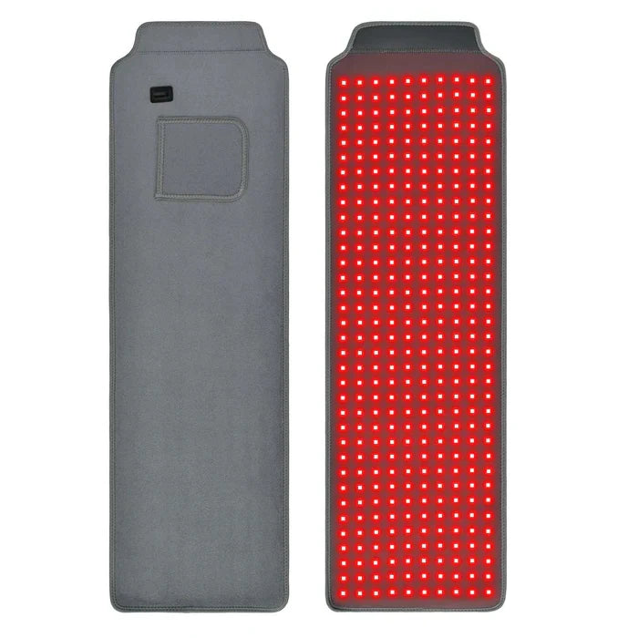
At home, the picture is messier. Brown University Health, Harvard Health, and Healthline all caution that consumer devices are usually less powerful and inconsistently specified. Many are cleared by the FDA primarily for safety, not for strong proof of efficacy for wound healing. The American Academy of Dermatology and UCLA Health recommend choosing devices that are explicitly FDA‑cleared for a relevant indication, following manufacturer instructions carefully, and consulting a dermatologist before using facial or high‑intensity devices, particularly in people with darker skin tones who are more prone to hyperpigmentation.
The scientific literature does, however, give you useful guardrails. For shallow wounds and early scars, I favor red wavelengths around 630–670 nm, because that is the range repeatedly used in fibroblast models, facial rejuvenation trials, and superficial wound studies. The 661 nm scratch assay indicates that doses near 3–4.5 J/cm² are a sweet spot for boosting fibroblast migration and proliferation without overshooting. The helium‑neon mouse work suggests that something on the order of 2 J/cm² can be effective in acute wounds, and the preclinical review of 68 studies points to 1–5 J/cm² as a common effective window.
Here is how this plays out practically. Suppose your device manufacturer actually lists an irradiance at a particular distance. If it is around 10 mW/cm² at the skin, a 5‑minute session would deliver roughly 3 J/cm² and an 8‑minute session about 4.8 J/cm², very similar to the most effective dose in the 661 nm fibroblast study. If the irradiance is closer to 20 mW/cm², those same doses would come at half the time. The Dior mask data show that higher fluences around 15.6 J/cm² can still be safe and beneficial for anti‑aging when used twice weekly, but the wound‑healing literature suggests starting in the lower, more conservative range and avoiding very frequent, long exposures in the same spot.
Equally important is spacing. The Dior mask trial, various PBM reviews, and my own experience all favor giving cells time to respond between sessions. Twice weekly with about 72 hours between exposures is a reasonable starting rhythm for cosmetic scars once the wound is clean, closed, and cleared by your clinician. For medically complex wounds like diabetic ulcers, be cautious: the best evidence involves supervised, protocolized light therapy in a clinic alongside aggressive standard care. That is not something to improvise with a beauty panel at home.
Finally, safety and context matter. Cleveland Clinic, Brown University Health, Healthline, Harvard Health, and the American Academy of Dermatology all highlight similar red‑light precautions. Avoid shining intense light directly into the eyes; use protective goggles with higher‑powered panels or facial masks. Get medical advice first if you are pregnant, have active cancer in the treatment area, a seizure history, a light‑sensitive condition like lupus, or take photosensitizing medications such as some antibiotics. People with darker skin tones and those prone to pigmentation issues should start with lower doses and involve a dermatologist, since visible light can sometimes darken spots when overused. And above all, do not let red light delay evidence‑based care for non‑healing, infected, or ischemic wounds.
FAQ
Can I use my red light beauty mask on a fresh surgical incision to prevent scars? The best data we have suggest red light works as an adjunct once your surgeon is happy with how the incision is closing and there are no signs of infection. Many studies that reported better scars or faster healing, such as those in sternotomy patients or hypertrophic scar children, applied light on clean, properly managed wounds and continued for weeks to months. Stanford and Harvard dermatology experts recommend asking your surgeon or dermatologist first, then using appropriately dosed red light in addition to, not instead of, scar‑care basics like sun protection, silicone, and follow‑up visits.
If red light is good, why not blast my wound with as much as possible? Because almost every serious review of photobiomodulation, including the 68‑study LASER/LED analysis and the 661 nm fibroblast work, shows a biphasic dose response. Very low doses do little; moderate doses help; high doses can flatten or even reverse the benefit. The preclinical helium‑neon study showed that multiplying a too‑low dose repeatedly did not catch up to a single, moderate optimum dose. Clinical summaries from Lumitex also warn that doses above roughly 50 J/cm² may be counterproductive. More is not better; better is better.
Will red light guarantee that I will not scar? No therapy can guarantee that, and any source that says otherwise is overselling. Genetics, wound size and depth, location, infection, tension on the wound, and systemic factors like diabetes or vascular disease all matter. What the current evidence suggests—from NASA‑initiated LED work, through burn and sternotomy trials, to modern dermatology reviews—is that appropriately dosed red and near‑infrared light can improve the odds: faster closure, more organized collagen, less chronic inflammation, and often softer, flatter, less noticeable scars. Think of it as a valuable tool in a broader toolkit that still includes meticulous wound care, smart lifestyle choices, and professional guidance.
Red light is not a miracle wand; it is a finely tunable signal to your biology. Treat it with the same respect you would a powerful medication, align it with the body’s own repair phases, and you can often persuade wounds to tell their story quietly, instead of carving it into your skin for life.
References
- https://lms-dev.api.berkeley.edu/red-light-therapy-research
- https://fcd.mcw.edu/?search/showPublication/id/43362
- https://www.health.harvard.edu/staying-healthy/red-light-therapy-for-skin-care
- https://epublications.marquette.edu/cgi/viewcontent.cgi?referer=&httpsredir=1&article=1249&context=dentistry_fac
- https://pubmed.ncbi.nlm.nih.gov/40175683/
- https://researcher.manipal.edu/en/publications/efficacy-of-multiple-exposure-with-low-level-he-ne-laser-dose-on-
- https://patients.sonoran.edu/treatment/cold-laser-and-led-light-therapy/
- https://news.usuhs.edu/2022/10/usu-professor-researches-novel-pain.html
- https://dspace.mit.edu/bitstream/handle/1721.1/104348/10103_2013_Article_1319.pdf?sequence=1&isAllowed=y
- https://med.stanford.edu/news/insights/2025/02/red-light-therapy-skin-hair-medical-clinics.html