
Hormonal Dermatitis: Where Hormones Meet Inflamed Skin
When people talk about “hormonal dermatitis,” they are usually describing a very real experience rather than a textbook diagnosis. The pattern is simple: whenever hormones swing—around the menstrual cycle, pregnancy, postpartum, perimenopause, or significant stress—skin that is already prone to dermatitis seems to revolt. Eczema patches thicken, redness spreads, itching spikes, and the barrier feels like it has simply given up.
Clinically, this sits under broader umbrellas like atopic dermatitis, seborrheic dermatitis, or other inflammatory rashes. The unifying biology remains the same: chronic inflammation, an impaired skin barrier, immune system overactivity, and, over time, microscopic damage to collagen and other structural proteins. Hormonal shifts do not change what the skin is made of; they change how reactive it becomes.
That is where red light therapy enters the conversation. The idea is compelling. If specific red and near‑infrared wavelengths can dial down inflammation, support wound healing, and stimulate collagen, could they help repair the damage that hormonal dermatitis leaves behind and maybe even make future flares less brutal?
As a long‑time light therapy geek who has tested more panels, masks, wands, and clinic devices than I care to admit, I want the answer to be yes. But the only honest way to approach this is to walk through what high‑quality dermatology sources, clinical trials, and eczema organizations actually show—and where the evidence is still thin or conflicting.

Red Light Therapy 101: What It Is and How It Acts on Skin
Red light therapy sits in a family of treatments often called photobiomodulation or low‑level light therapy. Unlike ultraviolet light, which can damage DNA and drive skin cancer, red and near‑infrared light use non‑UV wavelengths, typically around 600–700 nanometers for visible red and roughly 800–900 nanometers for near‑infrared.
According to dermatology centers and reviews cited by Cleveland Clinic, Stanford dermatology experts, and WebMD, these wavelengths are absorbed by mitochondria, the energy producers inside cells. That mitochondrial absorption appears to increase cellular energy (ATP), modulate oxidative stress, and trigger signaling pathways that affect fibroblasts, immune cells, endothelial cells, and keratinocytes. In plain language, the light nudges cells into a more “repair and recalibrate” mode rather than a “panic and inflame” mode.
Multiple sources, including Cleveland Clinic, Brown Health, and Dallas‑area dermatology practices, converge on similar core effects in skin:
Red and near‑infrared light can stimulate fibroblasts, increasing collagen and elastin production, which supports firmness and texture. They can reduce low‑grade inflammation and redness. They can improve blood flow and microcirculation, helping bring oxygen and nutrients to damaged tissue. They can support wound healing and, in some cases, reduce the appearance of scars.
In the formal language of a large phototherapy review indexed by the National Library of Medicine, photobiomodulation uses low‑level, non‑thermal light between roughly 600 and 1,300 nanometers to alter cellular function without ablation or injury. That is fundamentally different from ablative lasers or intense pulsed light, which deliberately damage tissue to trigger a wound‑and‑repair cascade.
How Red Light Differs From Classical Phototherapy for Eczema
Dermatologists have used light to treat skin disease for decades, but most of that history belongs to ultraviolet phototherapy. A comprehensive review of phototherapy in dermatology reports that both UVA and UVB, especially narrow‑band UVB, are well‑established, effective treatments for atopic dermatitis, with strong response rates in moderate to severe or chronic disease. Similar UV‑based protocols are used for psoriasis, vitiligo, and several rare dermatoses.
Red light therapy is not that. It does not use UVB or UVA, and it does not carry the same sunburn or skin‑cancer risk profile. It also does not yet have the same depth of evidence. For hormonal dermatitis and eczema‑like conditions, you can think of UV phototherapy as the established, prescription‑only, high‑powered option and red light as the gentler, lower‑risk but much less proven cousin.
That distinction matters when you are deciding what to trust for repairing real skin damage.

What the Research Actually Shows for Skin Repair
Anti‑Aging, Collagen, and Barrier Support
Before we zoom in on dermatitis, it is worth asking a simple question: can red light repair human skin at all?
A controlled randomized trial with more than one hundred volunteers looked at red and red‑plus‑near‑infrared light applied to the face two times per week for about fifteen weeks. Devices delivered non‑thermal, polychromatic light in the red band, with per‑session doses in the range of roughly 8 to 10 joules per square centimeter in that red spectrum. Compared with untreated controls, participants receiving red light showed measurable improvements in intradermal collagen density and reductions in skin roughness and wrinkle severity. Blinded physician assessors using standardized photographs confirmed a meaningful improvement in fine lines and texture. Importantly, the trial found that the narrower red‑only spectrum performed just as well as a broader red‑plus‑near‑infrared range at similar red doses.
Dermatology clinics echo those findings in real‑world practice. Multiple dermatologists interviewed for patient‑facing articles describe modest but noticeable improvements in fine lines, elasticity, and “glow” when patients commit to consistent red light sessions over several months. One dermatologist rated red light’s glow‑up potential about three and a half out of five, emphasizing that it works best as maintenance and a gentle adjunct, not a replacement for injectables or resurfacing.
For hormonal dermatitis, those anti‑aging and texture benefits matter because chronic inflammation and scratching degrade collagen and compromise barrier integrity over years. If red light can help rebuild collagen and improve barrier resilience, it theoretically makes the skin less fragile going into each hormonal swing, even if it is not controlling hormones themselves.
Wound Healing, Scars, and Post‑Procedure Recovery
Hormonal dermatitis often leaves marks: excoriations from scratching, thickened lichenified plaques, post‑inflammatory hyperpigmentation, and even scars from repeated trauma. Several sources, including Dallas and West dermatology practices, WebMD, and specialty recovery centers, highlight red light’s role in wound healing and scar modulation.
In wound settings, low‑level red and near‑infrared light can enhance blood flow, increase oxygen and nutrient delivery, and stimulate cellular proliferation. Observational data and early trials show that surgical incisions, minor injuries, and some burn wounds can close faster under structured light protocols, with better early scar quality. A phototherapy review notes that LED‑based light in the 660 to 904 nanometer range can improve wound healing parameters in chronic and surgical wounds over several weeks.
However, the data are mixed and nuanced. Stanford dermatology experts point out that in eyelid surgery studies, one trial found only a slight, non‑statistically significant edge for red‑treated areas, while another suggested faster early healing that leveled out by around six weeks, leaving treated and untreated sides looking similar. That pattern shows up often in photobiomodulation research: early speed‑ups that may or may not translate into long‑term cosmetic differences.
For dermatitis repair, that means red light might help calm and close the raw, over‑scratched areas more efficiently and could modestly improve how scars settle. It does not mean every scratch or plaque will magically disappear under a panel.
Eczema and “Hormonal Dermatitis” Flares
Now to the heart of the matter. Does red light specifically repair eczema‑like dermatitis, including when it is strongly influenced by hormones?
Medical News Today describes red light therapy as a form of phototherapy that may benefit eczema by reducing the inflammatory response and helping regulate immune overactivity. They note that while phototherapy overall has strong evidence in eczema, specific evidence for red light is still limited and emerging. The mechanism they highlight is important: red light can be absorbed by the skin and reduce oxidative stress, a recognized factor in eczema pathophysiology. A study published in 2024 reported that red light influenced inflammatory processes and modulated pro‑inflammatory cytokines, the chemical messengers that drive many immune reactions in skin.
That same article lists several skin‑focused benefits of red light with direct relevance to dermatitis repair: enhancing new skin cell growth, aiding wound healing, supporting scar management, rejuvenating the skin, and stimulating collagen and elastin. Together, those effects could help restore a damaged skin barrier over time, which is central to any eczema or dermatitis management strategy.
The National Eczema Association offers a more cautious view when it comes to consumer LED face masks and wands. They emphasize that LED therapy is not a standard‑of‑care eczema treatment. A small controlled study using blue light three times per week for four weeks in mild to moderate eczema showed improvement without adverse events, but experts stress that there are simply not enough clinical trials to endorse LED (red or blue) as a primary therapy.
Their guidance is practical and highly relevant for hormonal dermatitis. They advise against using LED devices on very inflamed or actively flaring eczema because even gentle LEDs can trigger irritation, redness, or dryness when the barrier is already severely compromised. During remission or quieter periods, they acknowledge that red and near‑infrared LEDs may offer subtle reductions in redness and irritation and may support barrier function, but they frame these as complementary benefits that sit alongside, not instead of, moisturizers, topical medications, and other prescribed therapies.
A separate evidence review summarized by ZOE takes an even stricter position: as of now, there is no good evidence that red light therapy can treat eczema as a primary disease‑modifying intervention. They note that some clinics still use red light based on its anti‑inflammatory potential and low side‑effect profile, but they underscore that the trials we would want—large, well‑controlled, eczema‑specific red light studies—are not yet in place.
Put those viewpoints together and a coherent picture emerges. For eczema and hormonal dermatitis patterns, red light therapy:
Has plausible mechanisms to help the skin recover between flares by reducing oxidative stress, modulating inflammatory signaling, and supporting barrier rebuilding via collagen and new keratinocyte growth. Has at least one emerging study suggesting it can influence inflammatory processes relevant to eczema. Has supportive but indirect evidence from wound healing and anti‑aging trials that show improved texture and collagen. Lacks strong, high‑certainty, eczema‑specific clinical trial data and is not considered a core, standalone treatment by major eczema organizations.
In other words, it can be a thoughtful adjunct for repair and maintenance in the right patient and the right phase, especially between hormonal flares, but it is not yet a proven eczema cure or a replacement for standard care.
Psoriasis and Other Inflammatory Dermatoses
Psoriasis is not hormonal dermatitis, but it shares features of chronic immune overactivation and barrier disruption. That makes psoriasis data useful for understanding how inflammatory dermatoses in general may respond to red light.
The broad phototherapy review notes that blue light has shown benefit in mild psoriasis vulgaris, improving keratinocyte behavior without cytotoxic damage. For psoriatic lesions in adults and children, narrow‑band UVB remains a mainstay when topical and systemic options are insufficient or contraindicated.
ZOE’s summary of red light research in psoriasis notes that the condition affects roughly 2 to 3 percent of the global population and that early red light results are “encouraging” but sparse. Some authors suggest blue light may be more effective than red for psoriasis plaques. That matters because many hormonal flares can aggravate both eczema‑like and psoriasis‑like tendencies in the same person.
The take‑home for hormonal dermatitis is that red light sits in a broader ecosystem of light‑based tools for inflammatory skin disease. UVB and UVA are proven but carry higher risk and require medical supervision. Blue and red LEDs are gentler and increasingly accessible but have less robust data. They may be worth layering in as adjuncts rather than as replacements for therapies that have decades of controlled data behind them.
Acne and Redness as Inflammation Models
Why talk about acne and rosacea in a dermatitis article? Because the way red light behaves in those conditions is one of the best real‑world windows into its anti‑inflammatory potential.
Dermatology practices and Harvard Health note that red LED light is commonly used to calm acne and photoaging changes. Blue light targets acne‑causing bacteria and oil glands, while red light targets inflammation and redness. Studies combining the two wavelengths show greater improvement in acne lesions compared with either one alone, suggesting that red light’s most reliable job is calming the inflammatory aftermath rather than eliminating the trigger itself.
Mayo Clinic dermatologists describe red light as one of several clinic‑based tools for acne and photoaging, with treatments delivered under prescription‑grade supervision. Over‑the‑counter masks and wands, by contrast, vary widely in wavelength, power, and safety. One early acne mask was recalled over concerns about potential eye damage in users with certain underlying conditions, emphasizing that even “gentle” LEDs are not toy‑level devices.
From an inflammation standpoint, those acne and rosacea experiences support the idea that red light is quite good at dialing down redness and low‑grade swelling. For hormonal dermatitis, that is precisely what you often want between flares: a way to reduce chronic background inflammation and give the barrier a calmer baseline from which to respond to hormonal swings.
Red Light vs Medical Phototherapy in Dermatitis Care
When you put red light therapy next to classic UV phototherapy, the contrast is stark.
Narrow‑band UVB and tailored UVA regimens have strong evidence in atopic dermatitis. In large pooled analyses of hundreds of patients, both UVA and UVB, particularly narrow‑band UVB, were identified as among the safest and most effective options for moderate to severe or chronic lichenified eczema when topical or systemic therapies are limited or contraindicated. UV‑based phototherapy is also effective in other rare dermatoses and conditions like cutaneous T‑cell lymphoma and vitiligo.
Red light therapy, by contrast, has robust evidence for cosmetic rejuvenation and hair growth, modest but real data for wound healing, and only early, mixed data for inflammatory dermatoses like eczema and psoriasis. It carries a much lower risk of DNA damage and sunburn because it avoids UV, but the trade‑off is that its disease‑modifying power is not as well demonstrated.
For hormonal dermatitis repair, the practical implication is this: if your dermatitis is severe, chronically debilitating, or complicated by infections, UV‑based phototherapy under a dermatologist’s supervision is still the better‑validated light option. Red light can be layered in around that, or used as a gentler tool when UV is not appropriate, but it should not delay or replace therapies with stronger evidence.

How I Would Integrate Red Light into a Hormonal Dermatitis Plan
This is where the “light therapy geek” mindset kicks in. When I evaluate red light for hormonal dermatitis repair, I am not asking whether it is a miracle. I am asking how to structure it so that it realistically supports barrier repair and inflammation control without overpromising.
Who Might Consider It
Based on the patterns described by Cleveland Clinic, Brown Health, National Eczema Association, and multiple dermatologists, red light therapy makes the most sense in a few scenarios.
It is plausible for someone whose dermatitis is reasonably controlled with moisturizers and medications but whose skin looks dull, rough, or scarred between hormonal flares. It is an option for people who cannot or prefer not to use more aggressive cosmetic interventions such as ablative lasers or harsh topicals, especially during pregnancy, when many anti‑aging drugs are off‑limits and dermatologists sometimes recommend red light as one of the few gentler cosmetic tools, with physician oversight. It can be considered for people who have already optimized their eczema regimen and want a low‑risk adjunct to support barrier resilience, knowing that benefits will likely be modest and gradual.
It is not a good fit for someone in the middle of a raging, open, weeping flare, for someone with known photosensitive conditions like lupus, or for anyone on highly photosensitizing medications unless a dermatologist has explicitly cleared it.
Dosing Sessions Based on Existing Protocols
Because high‑quality hormonal dermatitis trials do not exist yet, the only honest way to think about dosing is to look at how dermatologists and clinical trials use red light for nearby goals and adopt a conservative version of those patterns.
A dermatologist interviewed in a professional article cited in the research notes described in‑office facial red light sessions of about twenty minutes, three times per week, with cosmetic improvements accruing over three to six months. Other clinical and consumer‑facing sources, including West Dermatology, Brown Health, and WebMD, commonly describe protocols of ten to twenty minutes per session, performed several times per week for four to twelve weeks, with maintenance thereafter.
The anti‑aging randomized trial used twice‑weekly exposures for thirty sessions over about fifteen weeks, at power densities delivering around 8 to 10 joules per square centimeter in the red band. Participants did not experience serious adverse events, and tolerability was high.
Translating that into a hormonal dermatitis context, a cautious, repair‑oriented structure would look something like this in practice. You begin by having your dermatologist confirm the diagnosis and approve light therapy as an adjunct. You then patch‑test a small, less visible area—behind the ear or along the jawline—with a short exposure of just a minute or two, using eye protection, and wait a full day to watch for increased redness, burning, or delayed irritation. If the skin tolerates that, you gradually extend to sessions in the ten‑minute range, two or three times per week, always on relatively quiet skin rather than on actively oozing or severely inflamed plaques. Over eight to twelve weeks, you watch for changes in baseline redness, texture, and recovery time after hormonal flares.
The key is that you are not chasing daily exposure or the highest possible intensity; you are chasing consistency and tolerance. Multiple sources, including Stanford experts and Brown Health, emphasize that more light is not always better and that overtreatment can cause irritation or even blistering with powerful devices.
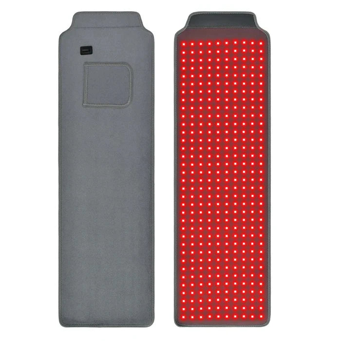
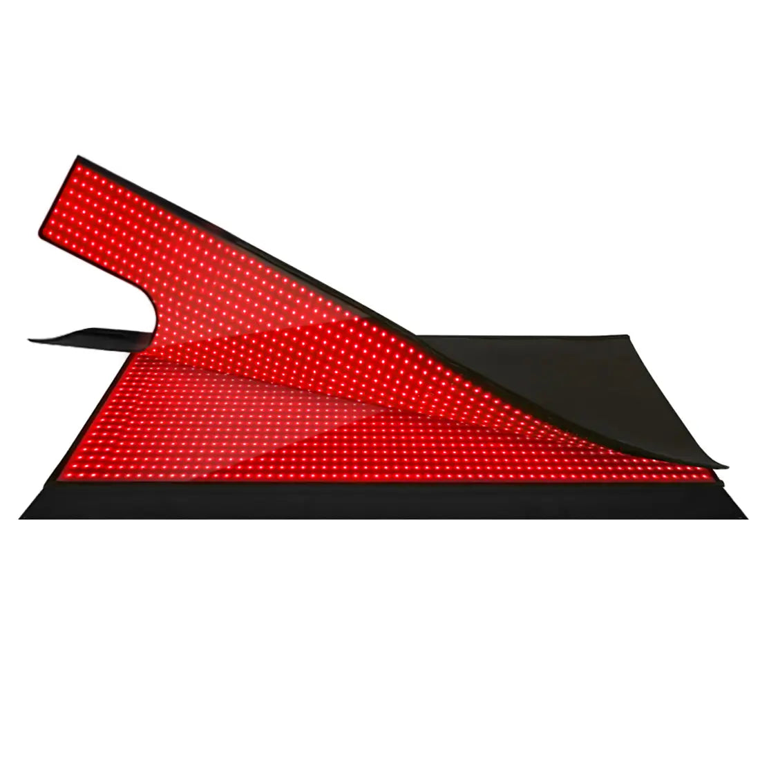
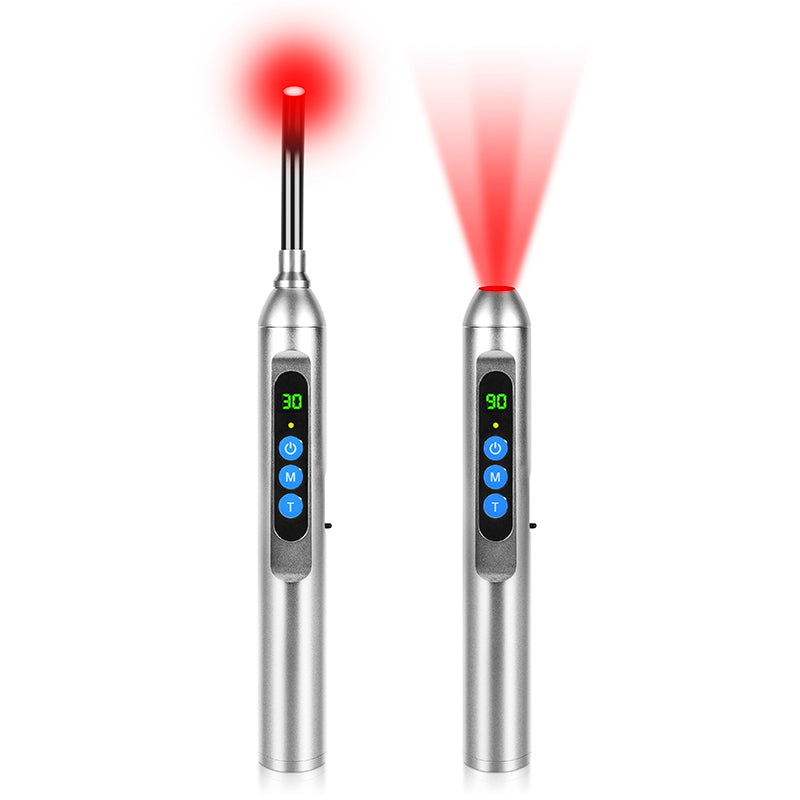
Device Choices: Clinic vs At‑Home
Device choice is where an experienced eye makes a huge difference.
Clinic‑based devices used by dermatologists and some integrative centers tend to have better‑characterized wavelengths and power densities. In the clinical trial mentioned earlier, irradiance and dose were precisely measured, and the devices emitted almost no erythemogenic UV radiation, making them safer from a sunburn perspective. For someone rehabbing hormonal dermatitis with significant scarring or post‑procedure changes, this kind of controlled setting offers the most predictable experience.
At the same time, FDA‑cleared at‑home panels, masks, and wands have made photobiomodulation more accessible. Cleveland Clinic and the National Eczema Association both point out that at‑home devices are often less powerful than in‑office equipment, which can lower risk but can also mean slower or smaller results. They recommend choosing devices that are cleared by the Food and Drug Administration for specific uses rather than unregulated gadgets that simply market “anti‑aging” or “eczema repair” without any safety review.
For sensitive or eczematous skin, the National Eczema Association notes that full‑face masks provide even coverage but may unnecessarily expose unaffected or very reactive areas. Wands and targeted panels allow you to confine treatment to zones that most need repair, which is often preferable when your dermatitis flares patchily along the jawline, neck, or hands.
Mayo Clinic and Harvard Health both stress the importance of eye protection for any facial treatment, even with consumer devices, because the long‑term safety of repeated eye exposure to intense LEDs is not fully known. A recall of a popular acne mask over eye‑safety concerns underscores that this is not a theoretical risk.
Safety, Risks, and Red Flags
Across Cleveland Clinic, WebMD, Brown Health, Stanford dermatology, and multiple clinic articles, the safety profile of red light therapy for skin is generally favorable when devices are used appropriately. Treatments are non‑invasive, non‑UV, and usually painless, with side effects limited to temporary redness, warmth, or mild irritation in most users.
However, there are several important caveats if you are thinking specifically about hormonal dermatitis repair.
Long‑term safety data for daily or near‑daily home use over many years are still limited. Most studies run for weeks to a few months. Experts repeatedly caution that we do not yet know the full lifetime impact of chronic LED exposure on skin and eyes. Misuse can cause harm. Very high doses or excessively long sessions on high‑powered panels have produced skin redness and blistering in early trials. Shining aggressive light directly into the eyes can damage the retina, especially in people with underlying eye disease or those on medications that increase light sensitivity. Certain populations require special caution. People with photosensitive conditions such as lupus, those with a history of skin cancer, and those taking photosensitizing medications should not start red light on their own. Evidence in pregnancy is limited; one large laser‑based study suggests safety, but most medical centers still recommend that pregnant patients clear any light‑based treatment with an obstetric provider and dermatologist. Device quality is highly variable. Harvard Health notes that the Food and Drug Administration reviews many consumer devices for safety, not efficacy, so a device can be “cleared” without strong proof that it does what marketing promises. Off‑brand or unregulated devices may have inaccurate wavelength claims, uneven output, or general quality issues. Overconfidence can delay effective care. Perhaps the biggest red flag is using red light therapy as a substitute for seeing a dermatologist about new or changing lesions. Multiple experts explicitly warn against treating “sun damage” or chronic rashes at home without first confirming that the skin changes are not cancer, infection, or another condition that will not respond to red light.
For hormonal dermatitis specifically, the safest path is to think of red light therapy as a supportive tool layered onto a rock‑solid foundation: diagnosis, trigger management, barrier‑repair routines, prescribed topicals when needed, and, for some, systemic therapies. Red light can help the skin do its job better between hormonal storms, but it is not a shield against those storms.

A Snapshot of the Evidence for Hormonal Dermatitis‑Relevant Goals
Goal or Condition |
Evidence for Red Light Therapy |
Likely Role Today |
Key Caveats |
Repairing photoaged, rough, or dull skin |
Controlled trials show improved collagen, wrinkles, and roughness |
Valid cosmetic adjunct for texture and “glow” |
Effects are modest and develop over months; not a substitute for lasers |
Healing scratches, minor wounds, and scars |
Early trials and clinical reports show faster early healing |
Reasonable adjunct for post‑procedure and minor trauma |
Long‑term cosmetic differences may be small; protocols vary widely |
Eczema and hormonal dermatitis repair |
Mechanistic and early data, but no high‑certainty eczema trials |
Experimental adjunct for barrier support between flares |
Not standard of care; major eczema groups urge caution and realistic goals |
Psoriasis and related dermatoses |
Encouraging but sparse data; blue light appears more effective |
Possible adjunct when UV phototherapy is unsuitable |
UV‑based phototherapy remains far better studied and more potent |
Acne, redness, and low‑grade inflammation |
Multiple studies support combined red and blue light |
Well‑accepted adjunct for inflammatory acne and redness |
Does not replace root‑cause acne treatments or medical evaluation |
Brief FAQ
Can red light therapy stop hormonal dermatitis flares from happening?
There is no good evidence that red light can prevent hormonally driven flares altogether. The more realistic aim is to help the skin recover better between flares by improving barrier resilience and calming background inflammation, which may make each flare less destructive to the skin over time.
Is it safe to use a red light mask on my face if I have eczema?
Major eczema organizations consider LED masks generally safe for many skin types but advise caution for eczema. They recommend avoiding masks on very inflamed, actively flaring skin; starting with a short patch test on a small area; using them mainly during remission or quieter periods; and always discussing the plan with a board‑certified dermatologist, especially if your eczema is moderate to severe.
How long before I see any benefit for dermatitis‑related damage?
For cosmetic changes like smoother texture and fine wrinkles, clinical and clinic reports usually describe visible changes over about three to six months of consistent use. For dermatitis repair, you are watching a slower game: do plaques heal more cleanly, does redness between flares decrease, and does your barrier feel more resilient season to season. Those shifts, if they happen, tend to be gradual rather than dramatic.

Closing Thoughts from a Light Therapy Geek
Red light therapy absolutely changes skin biology; that much is clear from controlled trials and decades of photobiomodulation research. Where the science is still catching up is in telling us exactly how far that change goes for complex, hormone‑sensitive dermatitis. If you treat red light as a precise, gentle tool to support barrier repair and collagen—not as a hormonal reset button—and you combine it with smart dermatologic care, it can earn a spot in your recovery arsenal. Respect the limits of the evidence, protect your eyes and your expectations, and let the data, not the marketing, steer how you shine that light on your skin.
References
- https://www.health.harvard.edu/diseases-and-conditions/led-lights-are-they-a-cure-for-your-skin-woes
- https://digitalcommons.kansascity.edu/cgi/viewcontent.cgi?article=2017&context=studentpub
- https://pmc.ncbi.nlm.nih.gov/articles/PMC3926176/
- https://dev.ppc.uiowa.edu/libweb/4P8037/HomePages/LightTherapyForAllergies.pdf
- https://med.stanford.edu/news/insights/2025/02/red-light-therapy-skin-hair-medical-clinics.html
- https://soar.suny.edu/bitstream/handle/20.500.12648/15752/Austin_as_entered_20210816.pdf?sequence=1&isAllowed=y
- https://do-server1.sfs.uwm.edu/slug/365M95686G/text/145M32G/the_science-of__phototherapy.pdf
- https://www.brownhealth.org/be-well/red-light-therapy-benefits-safety-and-things-know
- https://my.clevelandclinic.org/health/articles/22114-red-light-therapy
- https://newsnetwork.mayoclinic.org/discussion/mayo-clinic-minute-dermatologist-explains-light-therapy-for-skin/