
Red light therapy has gone from NASA lab curiosity to a staple in high-end dermatology clinics and serious home biohacking setups. Yet most people using a red light panel on their face, joints, or whole body could not tell you what is actually changing inside a single cell when those photons land.
As someone who obsesses over light dosing, wavelengths, and real-world outcomes, I can tell you this: red and near-infrared light do not work by “mystical energy.” They work because they drive very specific biochemical reactions. When you understand those reactions, you can design safer, smarter protocols instead of chasing hype.
In this article, we will unpack seven key cellular chemical reactions triggered by red and near-infrared light, grounded in current research from major medical centers, peer‑reviewed journals, and clinical trials. You will also see where the evidence is strong, where it is speculative, and how to translate these mechanisms into practical use at home or in a clinic.
Red Light Therapy In A Nutshell
Red light therapy, also called photobiomodulation or low-level light therapy, uses low-intensity red and near‑infrared wavelengths, roughly in the 600–1100 nanometer range. These wavelengths are delivered by LEDs or low‑power lasers to skin and underlying tissue without heating or burning it.
NASA originally explored red LEDs to grow plants and help astronauts heal wounds more quickly. Since then, red light has been integrated into medical and wellness practice for skin rejuvenation, wound healing, pain, and hair loss. Dermatology and cancer centers use it both as a stand‑alone photobiomodulation treatment and as the light component of photodynamic therapy, where a photosensitizing drug plus red light selectively destroys abnormal cells.
A Stanford dermatology review, Cleveland Clinic, Harvard dermatology, and MD Anderson Cancer Center all describe red light therapy as non‑invasive, non‑UV, and generally safe when used properly, with the strongest evidence today for hair growth, some skin rejuvenation, and certain forms of oral mucositis and pain. At the same time, these centers emphasize that many claims—especially for broad systemic disease and weight loss—are still unproven and require more rigorous trials.
With that context, let us zoom down to the seven core chemical reactions happening inside your cells when you flip the red light on.

Reaction 1: Photon-Driven ATP Production In Mitochondria
The first and most central reaction is a boost in cellular energy production.
Inside almost every cell sit mitochondria, the double‑membrane “power plants” that use nutrients and oxygen to produce adenosine triphosphate, or ATP. You recycle roughly your body weight in ATP every day, according to cellular respiration research summarized by Joovv and others. Most of that ATP comes from aerobic respiration: a coordinated series of reactions that strip electrons from food, pass them down the electron transport chain, pump protons, and drive ATP synthase.
Red and near‑infrared photons are absorbed by cytochrome c oxidase, the terminal enzyme of that electron transport chain. A mechanistic review on low‑intensity light therapy identifies cytochrome c oxidase as a key photoacceptor, with action spectrum peaks in the red and near‑infrared bands around 613–623, 667–684, 750–772, and 812–846 nanometers. When cytochrome c oxidase absorbs light in those bands, it passes electrons more efficiently, increases proton pumping, and ultimately drives more ATP synthesis.
You can think of the core chemical shift this way: oxidized components in the respiratory chain accept electrons faster after absorbing photons, so the conversion of oxygen, water, and nutrients into ATP accelerates. Several sources, including Joovv’s cellular respiration explainer and mechanistic reviews, note that red and near‑infrared exposure can also increase the number of mitochondria over time and improve their overall performance.
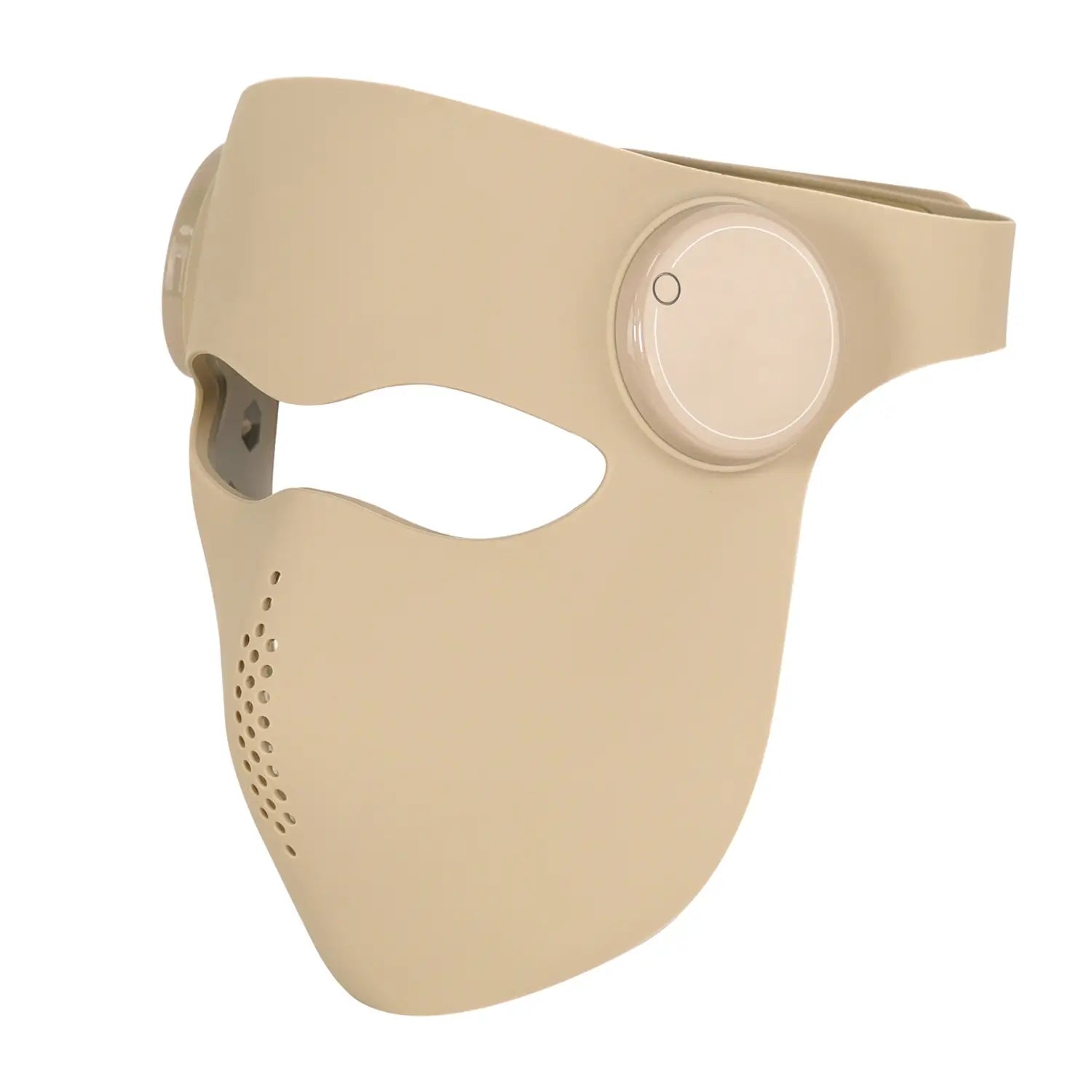
A concrete human example comes from a French clinical trial using a home‑use red LED mask at about 630 nanometers. The mask delivered an average irradiance of 21.7 milliwatts per square centimeter and a dose of 15.6 joules per square centimeter during each twelve‑minute session. Participants, aged 45 to 70, used it twice a week for three months. Researchers measured wrinkle depth, skin firmness, elasticity, dermal density, and complexion. The results showed progressive improvements over one, two, and three months, and those gains persisted for up to fourteen to twenty‑eight days after stopping treatment. That persistence suggests structural changes driven by altered ATP-dependent repair processes, not just a transient fluid shift.
From a practical standpoint, this ATP reaction explains why red light is used for skin rejuvenation, wound healing, muscle recovery, and even certain neurological applications. Higher ATP availability means cells have more energy for repair, signaling, and detoxification. The flip side is the dosing problem: too little light does nothing, while too much can inhibit function, a pattern known as the biphasic dose response or Arndt–Schulz law. The French mask study intentionally spaced sessions seventy‑two hours apart to respect what the investigators called “cellular energy digestion time,” highlighting that more sessions are not automatically better.

Reaction 2: Nitric Oxide Photodissociation And Vascular Signaling
The second key reaction involves nitric oxide, a small gaseous signaling molecule with outsized effects on both mitochondria and blood vessels.
Nitric oxide can bind to cytochrome c oxidase and compete with oxygen at its active site. When that happens, electron transport slows, ATP production drops, and reactive oxygen species can accumulate. Mechanistic work summarized by Joovv, Carolina Functional Neurology Center, and redox-focused reviews describes nitric oxide binding to cytochrome c oxidase as a reversible brake on respiration.
Red and near‑infrared photons can dislodge that nitric oxide from the enzyme. In biochemical terms, light energy helps break the bond between nitric oxide and the copper centers of cytochrome c oxidase, freeing the enzyme to bind oxygen again and resume efficient catalysis. The nitric oxide released this way does not just vanish; it diffuses and can relax smooth muscle in blood vessel walls, promoting vasodilation.
A Nature commentary on red and near‑infrared light as a countermeasure for spaceflight‑associated neuro‑ocular syndrome notes that this dissociation of nitric oxide, coupled with improved mitochondrial intermembrane potential and ATP production, is one of the hypothesized benefits of red and near‑infrared exposure for stressed retinal tissue.
Clinically, this nitric oxide reaction is reflected in several observations:
Dermatology experts at Stanford and Harvard describe red light–induced vasodilation as one reason hair follicles receive more oxygen and nutrients, helping regrow thinning hair when treatment is consistent for months. They also note that benefits stop when therapy stops and that dead follicles cannot be revived, which fits a model where improved blood flow and mitochondrial function rescue struggling but not lost cells.
Pain and rehabilitation centers, including MD Anderson Cancer Center and multiple wellness clinics, point out that red light helps reduce pain and inflammation in joints and muscles partly by improving local circulation. Better perfusion clears inflammatory mediators and delivers more oxygen and nutrients to tissues in need of repair.
Some wellness‑oriented sources go further, suggesting that this nitric‑oxide‑mediated vasodilation supports rheumatoid arthritis, slow‑healing wounds, and cardiovascular conditions, and may even help insulin resistance. The mechanistic logic is plausible, but large, controlled human trials in those areas remain limited, and major medical organizations are cautious about making strong claims.
There is also an important caveat. A scientific review on red and near‑infrared–stimulated angiogenesis highlights that nitric oxide and vascular endothelial growth factor are major drivers of new blood vessel growth. In healthy or injured tissues, that is usually good news. In tumors, where nitric oxide and vascular endothelial growth factor are already promoting pathological angiogenesis, additional pro‑angiogenic signaling could potentially be harmful. That is why most oncology groups recommend discussing any light‑based therapy with an oncologist before using it around known or suspected cancers.
Reaction 3: Transient Reactive Oxygen Species Bursts And Redox Signaling
The third reaction revolves around reactive oxygen species, or ROS. These are chemically reactive oxygen‑containing molecules such as superoxide and hydrogen peroxide that are generated during normal metabolism.
A detailed redox‑mechanism review of low‑intensity light therapy defines cellular redox state as the balance between reactive oxygen species production and antioxidant removal. High levels of reactive oxygen species are cytotoxic and can damage lipids, proteins, and DNA. Low levels, in contrast, act as signaling molecules that regulate growth, differentiation, and stress responses.
When cytochrome c oxidase absorbs red or near‑infrared photons and accelerates electron transport, it does not simply increase ATP. It also generates a short‑lived, low‑level burst of reactive oxygen species. That transient spike shifts redox‑sensitive signaling pathways, including changes in pH and in intracellular calcium, and activates transcription factors that drive gene expression.
Experiments with human skin fibroblasts exposed to red and near‑infrared light at wavelengths like 632.8, 812, and 820 nanometers show exactly this pattern: a temporary increase in reactive oxygen species correlates with increased cell proliferation. Microarray analyses have reported that red light can modulate expression of more than one hundred genes, mostly enhancing proliferation and suppressing programmed cell death. These gene expression changes underlie macroscopic effects such as faster wound closure and improved tissue repair.
Wellness‑oriented summaries often say that red light therapy “reduces oxidative stress,” which sounds like it simply scavenges reactive oxygen species. The mechanistic work suggests a more nuanced picture. The therapy appears to use a small, controlled reactive oxygen species pulse as a signal to upregulate antioxidant defenses and repair machinery, restoring a healthier balance over time. The therapeutic window is therefore crucial. The redox review emphasizes that effective photobiomodulation depends critically on wavelength, dose, intensity, and even pulse frequency, with the goal of eliciting brief, low‑level redox signals rather than sustained oxidative stress.
Clinically, this redox balancing act is particularly relevant for conditions marked by chronic oxidative stress, such as diabetic wounds and chronically inflamed joints. Proliferating cells, diabetic wounds, and inflamed tissues often exhibit pro‑oxidant redox states and seem especially responsive to low‑intensity light. That is one reason red and near‑infrared therapy is being explored for slow‑healing diabetic ulcers and tendinopathies. However, as Cleveland Clinic and WebMD both caution, many of these indications rely on small or moderate‑quality studies, and standard‑of‑care treatments should not be abandoned in favor of red light alone.
Reaction 4: Calcium Ion Waves And Membrane Depolarization
The fourth reaction is less famous in popular discussions but critical for understanding red light’s effects on nerves, brain, and blood vessels: changes in calcium ion dynamics.
Calcium ions are one of the body’s main intracellular messengers. They regulate neurotransmitter release, gene transcription, muscle contraction, and even cell death. A series of experiments studying photobiomodulation in neurons and cancer cells has shown that low‑intensity red and near‑infrared light, especially around 650 and 808 nanometers, can increase calcium influx across the cell membrane without heating the tissue.
In those experiments, red and near‑infrared light changed the membrane potential, making the inside of the cell less negative, and opened calcium‑permeable channels, including ionotropic glutamate receptors. Calcium then flowed in from outside the cell. In addition, light triggered calcium release from the endoplasmic reticulum, the major intracellular calcium store, and some of that calcium was taken up by mitochondria.
In practical terms, this means that a burst of red or near‑infrared light can create a coordinated wave of calcium signaling: entry through membrane channels, release from internal stores, and uptake by mitochondria. At carefully chosen doses, this appears to stabilize overly stressed neurons, reduce endoplasmic reticulum stress, and promote repair. At excessive doses or in vulnerable cells, too much calcium can be harmful, contributing to excitotoxicity and cell death.
These calcium‑dependent mechanisms help explain why headsets and helmets delivering red or near‑infrared light through the skull and even intranasally are being studied for cognitive benefits. UCLA Health highlights early research in people with dementia where intranasal and transcranial red light therapy used for about six minutes a day over eight weeks produced measurable improvements in cognitive function without significant adverse effects. A review summarized by WebMD on dementia and red light found improvements in all ten included trials, though most were small and preliminary.
Neurorehabilitation reviews cited by Carolina Functional Neurology Center point to red and near‑infrared photobiomodulation as a promising strategy for conditions like traumatic brain injury, stroke, Parkinson’s disease, and depression, with mechanisms spanning mitochondrial support, reduced neuroinflammation, and modulation of apoptotic signaling. Calcium regulation likely intersects with all three.
For a serious user, the implication is clear. If you are targeting the brain, you are not just chasing “more ATP.” You are modulating calcium‑dependent signaling in neurons. That makes careful dosing, medical supervision, and realistic expectations essential. The same dose that helps an aging but otherwise stable brain may not be appropriate for someone with uncontrolled seizures or acute brain injury.

Reaction 5: Gene Expression, Collagen Building, And Stem Cell Activation
The fifth reaction is downstream of ATP, nitric oxide, reactive oxygen species, and calcium: changes in gene expression that drive new protein synthesis, especially collagen and elastin, and activation of stem and progenitor cells.
A mechanistic review of low‑intensity light therapy using microarray data found that red light modulated the expression of more than one hundred genes across several functional categories. Most changes favored proliferation and survival, including upregulation of genes involved in cell cycle progression and downregulation of genes promoting apoptosis. Mitochondrial networks also reorganized, another sign that the cell was shifting into a pro‑repair state.
In skin, fibroblasts are the cells responsible for producing collagen and elastin, the structural proteins that give skin its firmness and elasticity. A clinical trial on red and near‑infrared treatment for skin aging reported increased intradermal collagen density and reductions in wrinkles and skin roughness. Omaha Massage and Wellness Center, summarizing that work in Photomedicine and Laser Surgery, notes about a thirty‑one percent increase in collagen density after twelve weeks of treatment.
The French LED mask study mentioned earlier adds more detail. Volunteers continued their usual skin care while adding two twelve‑minute red light sessions a week for three months. Objective measurements showed improved dermal density, firmness, elasticity, and texture, and those improvements persisted for up to a month after stopping treatment. That durability indicates that gene expression and protein remodeling, not just temporary swelling, were at work.
Stem cells are also affected. Deeply Vital Medical, drawing from stem cell research, reports that red light can increase stem cell viability and survival, promote their migration toward sites of injury, and support differentiation into needed cell types. Most of these data focus on mesenchymal stem cells, which have strong regenerative potential in musculoskeletal and connective tissue. Combined with collagen synthesis, this creates a biochemical basis for why red light is used as an adjunct in joint and tendon rehabilitation.
The immune system is not left out. Reviews and wellness clinics describe red light as enhancing certain immune functions, in part by stimulating stem cells and changing growth factor and cytokine expression. Mechanistic angiogenesis research shows that red and near‑infrared light can alter macrophage behavior, boosting growth factor production and shifting cytokine profiles toward tissue repair.
There is, however, a boundary. Gene expression is not a precision tool in this context. Red light does not know the difference between a fibroblast that should build healthy dermis and a malignant cell in a tumor microenvironment. Experimental data on light’s effects on cancer cells and vascular endothelial growth factor in malignancy are mixed, with some wavelengths and doses stimulating proliferation and others inhibiting it. Until that picture is clearer, major cancer centers recommend using red light cautiously and under medical supervision in people with current or prior malignancies.

Reaction 6: Water Structuring And The Mitochondrial “Battery”
The sixth reaction sits at the edge of mainstream and emerging biophysics: changes in how water behaves near biological surfaces and how that relates to membrane potential.
Mitochondria generate ATP by maintaining a proton gradient across their inner membrane. Classic bioenergetics describes this as protons being pumped into the intermembrane space, creating an electrochemical gradient that drives ATP synthase. Recent work in cell biophysics, summarized in an accessible way by Recharge Health, adds another layer: the concept of exclusion zone water.
Exclusion zone water, sometimes called structured water, refers to a more ordered phase of water that forms next to hydrophilic surfaces, including membranes and proteins. This water is more viscous, contains fewer solutes, and carries a different charge distribution than bulk water. In laboratory models, red and near‑infrared light expand this exclusion zone, effectively increasing the region where water can store and separate charge.
According to Recharge Health’s mechanistic overview, red light increases the exclusion zone water inside cells and in mitochondria, enhancing the electrical potential across the mitochondrial membrane and improving the cell’s ability to store and use energy. This would mean that not only the classical proton gradient but also structured water layers participate in the mitochondrial “battery.”
Mainstream clinical literature on red light therapy does not yet routinely describe exclusion zone water, but it does report increased mitochondrial membrane potential after red and near‑infrared exposure, as in the Nature commentary on neuro‑ocular syndrome in astronauts. Whether part of that change is mediated by water structuring remains an open but intriguing question.
For the practical user, the important takeaway is less about the fine details and more about respecting the system. You are not simply shining light on a passive bag of fluid. You are interacting with a finely tuned electrochemical device composed of membranes, proteins, and water. This is another reason why protocols like the French mask trial avoid daily, high‑dose bombardment and instead favor moderate doses with rest days, allowing the system to integrate and stabilize the changes.
Reaction 7: Angiogenesis, Glucose Control, And Metabolic Tuning
The seventh reaction involves the vascular and metabolic systems: new blood vessel formation, blood flow changes, and shifts in glucose handling.
A mechanistic study on red and near‑infrared light–stimulated angiogenesis shows two main pathways at work. First, activation of mitochondrial cytochrome c oxidase increases ATP and reactive oxygen species, which in turn activate signaling cascades. Second, red and near‑infrared light can induce a rapid calcium influx and membrane depolarization even independent of cytochrome c oxidase. Together, the increased reactive oxygen species and cytosolic calcium alter protein conformation, gene transcription, and secretion of vascular endothelial growth factor and nitric oxide in endothelial cells.
Vascular endothelial growth factor and nitric oxide are central drivers of angiogenesis, the growth of new blood vessels. Under normal conditions, angiogenesis is largely quiescent. When tissue is injured or hypoxic, vascular endothelial growth factor rises, and new vessels sprout to restore oxygen and nutrient supply. Red and near‑infrared light, by increasing vascular endothelial growth factor production and nitric oxide synthesis in endothelial cells and by modulating macrophage behavior, can enhance this repair process. In wound models, red and near‑infrared light at wavelengths like 629–660, 780, and 804 nanometers have been shown to stimulate endothelial proliferation and formation of capillary‑like structures.
Clinically, this pro‑angiogenic reaction contributes to faster wound healing and early improvements in post‑surgical scars seen in some studies, even though long‑term scar differences may fade. It also likely supports the improved dermal density and skin perfusion reported in anti‑aging LED mask trials and in studies reviewed by Harvard dermatology and the American Academy of Dermatology.
Beyond blood vessels, red light also interacts with metabolism. A human trial from a major research university exposed participants’ backs to 670‑nanometer red light for fifteen minutes, then performed a standard oral glucose tolerance test. Compared with placebo, the red light group showed a twenty‑seven point seven percent reduction in overall blood glucose exposure and a seven point five percent lower peak glucose spike over the two‑hour test window. Mechanistically, the authors attributed this effect to enhanced mitochondrial ATP production increasing glucose uptake and consumption.
In animals, long‑wavelength light in the 650–900 nanometer range has been shown to improve mitochondrial function, reduce blood glucose, and extend health span. The human data are early but provide biochemical support for the idea that red and near‑infrared light can modulate systemic metabolism through local mitochondrial stimulation, a pattern sometimes described as an abscopal effect.
Wellness clinics and metabolism‑focused writers often extend this into weight‑loss claims, noting studies where red light appears to influence inflammatory markers, insulin signaling, and even appetite hormones like leptin and ghrelin. Deeply Vital Medical, for example, discusses red light’s potential to support lipolysis, lymphatic clearance of fat breakdown products, and better insulin sensitivity.
However, Cleveland Clinic is explicit that there is currently no solid scientific evidence that red light therapy alone produces meaningful weight loss, cellulite removal, or mental health treatment, despite widespread online marketing. A reasonable synthesis is that red and near‑infrared light may support healthier metabolic signaling and blood flow, especially when combined with diet, movement, and sleep improvements, but treating it as a fat‑melting shortcut is not supported by mainstream clinical data.
Once again, context matters. In tissues that need more blood flow and better glucose handling, these angiogenic and metabolic reactions are beneficial. In tumors or advanced atherosclerotic plaques already rich in pathological vessels and inflammatory signaling, indiscriminately amplifying vascular endothelial growth factor and nitric oxide may not be wise. That is why people with significant cardiovascular or oncologic histories should discuss red light plans with their physicians.
Quick View: Seven Cellular Reactions And What They Mean
Reaction |
Main molecules and processes |
Where evidence is strongest today |
Key opportunity |
Key caution |
Mitochondrial ATP boost |
Cytochrome c oxidase, electron transport, proton gradient, ATP |
Skin rejuvenation, wound healing, some pain conditions |
More cellular energy for repair and performance |
Biphasic dose; too much light can inhibit rather than help |
Nitric oxide photodissociation and vasodilation |
Nitric oxide release from cytochrome c oxidase and vessels |
Hair growth, local circulation, some pain relief |
Better microcirculation and oxygen delivery |
Pro‑angiogenic signaling may be risky in active cancers |
Transient reactive oxygen species and redox signaling |
Superoxide, hydrogen peroxide, antioxidant systems, redox sensors |
Chronic wounds, inflamed joints (early data) |
Low‑level stress signal that upregulates repair pathways |
Overdosing can drive oxidative damage rather than balance |
Calcium ion influx and membrane depolarization |
Calcium channels, endoplasmic reticulum release, mitochondria |
Experimental neuroprotection, early dementia studies |
Support for neuroplasticity, brain repair, and pain relief |
Mis‑dosed light could worsen excitotoxicity in some brains |
Gene expression and collagen, elastin, stem cells |
Transcription factors, collagen and elastin genes, stem cell genes |
Anti‑aging dermatology, joint and tendon support |
Structural tissue remodeling and regeneration |
Gene shifts are non‑specific in cancer‑prone environments |
Water structuring and membrane potential |
Exclusion zone water, proton gradients, membrane charge |
Mechanistic and biophysical models; indirect human data |
Potentially more stable cellular “battery” and resilience |
Still emerging science; not yet a primary clinical metric |
Angiogenesis and metabolic tuning |
Vascular endothelial growth factor, nitric oxide, glucose handling |
Wound healing, early glucose control, some pain conditions |
Better perfusion and possibly smoother glucose responses |
Angiogenesis in tumors or plaques could be undesirable |
How To Work With These Reactions In Real Life
The science is fascinating, but most people reading this are trying to decide how, or whether, to use red light therapy at home or with a practitioner.
Medical centers like Cleveland Clinic, Harvard dermatology, Stanford, UCLA, MD Anderson, and WebMD converge on a few practical themes.
First, treat red light as an adjunct, not a stand‑alone cure. For skin, it sits alongside sun protection, a sane skin care routine, sleep, and nutrition. For joints and muscles, it complements physical therapy, strength work, and, when needed, medications. For metabolic or neurological goals, it layers on top of the fundamentals: diet, movement, stress management, and mainstream medical care.
Second, pay attention to device quality and dosing. Professional devices in dermatology or pain clinics are more powerful and better characterized in terms of wavelength and output. At‑home devices—masks, caps, wands, and panels—are convenient but often have unclear or overstated specifications. When possible, choose products that specify wavelengths in the proven therapeutic windows, such as red around 630–670 nanometers and near‑infrared around 800–850 nanometers, and that provide measured irradiance so you can estimate dose.
Clinical protocols provide clues for reasonable dosing. The French LED mask trial achieved significant anti‑aging effects with just two twelve‑minute sessions per week for three months. Cleveland Clinic notes that many dermatologic protocols involve one to three sessions per week over weeks to months, sometimes followed by maintenance sessions. UCLA’s dementia study used six minutes a day for eight weeks. None of these protocols used hours of daily full‑body exposure at maximal intensity.
Third, respect safety boundaries. Red light devices do not emit ultraviolet light and have not been linked to skin cancer. Short‑term side effects are usually limited to mild redness, and even pregnant women have been exposed to low‑level laser treatments without obvious short‑term harm in at least one study. That said, misused devices can cause skin damage or burns, and direct eye exposure to intense light can injure the retina. Clinics typically use goggles or shields, and home users should follow instructions and protect their eyes, especially with facial panels or masks.
People with light‑sensitive conditions such as lupus, those taking photosensitizing medications, and individuals with a history of skin cancer, serious eye disease, or active malignancy should consult physicians before starting red light protocols. Dermatology associations also advise that people with darker skin tones start cautiously and under professional guidance, since visible light can sometimes worsen hyperpigmentation in sensitive skin.
Finally, remember costs and commitment. Red light sessions in clinics and spas are often not covered by insurance and can become expensive over months of regular visits. At‑home devices require an upfront investment that can run into hundreds of dollars or more. The benefits, where they exist, tend to build gradually over weeks or months and fade when treatment stops. That makes red light therapy best suited for people willing to use it consistently and pair it with lifestyle changes, not for those hoping for a one‑time fix.
FAQ For The Serious Light Therapy Geek
Is daily red light therapy better than a few times per week?
Not necessarily. Mechanistic reviews on photobiomodulation and the clinical LED mask study in France both emphasize a biphasic dose response: too little light has no effect, and too much can inhibit cellular function or drive unhelpful stress. The mask protocol used two twelve‑minute sessions per week and still produced clear, measurable improvements in skin structure that lasted up to a month after stopping. Many clinical protocols for skin and pain cluster around short sessions a few times per week rather than daily high‑intensity exposure. If you are using a home device, starting at manufacturer‑recommended times a few days a week and reassessing with your clinician is more evidence‑aligned than simply using it as long and as often as possible.
Can red light therapy really help with weight loss?
Metabolism‑focused clinics and some research groups highlight that red and near‑infrared light can improve mitochondrial ATP production, modulate inflammation, and even influence insulin signaling and appetite hormones. A human study using 670‑nanometer light showed a meaningful reduction in blood glucose response after a glucose drink, which is promising for metabolic health. However, Cleveland Clinic explicitly notes that there is no strong scientific evidence that red light therapy alone produces sustainable weight loss or cellulite removal. The fairest reading today is that red light may become a useful adjunct for metabolic health—especially when paired with diet, movement, and sleep—and that marketing it as a fat‑melting shortcut is not justified by high‑quality data.
How should a serious biohacker choose and use a device?
If you care about mechanisms, look first at wavelength and power density. Panels or masks that clearly state red wavelengths around 630–670 nanometers and near‑infrared around 800–850 nanometers align well with action spectra for cytochrome c oxidase and with the devices used in many studies. Seek brands that provide measured irradiance rather than vague “high power” claims, and be wary of gadgets that do not specify outputs at all. Consider your primary target: for scalp or facial skin, a mask, cap, or smaller panel may suffice; for joints or larger muscle groups, a panel with good coverage is more practical. Then design a protocol that mirrors clinical patterns—short sessions, a few times per week, with periodic re‑evaluation—rather than ad‑hoc blasts of light whenever you remember.
Stepping back, the magic of red light therapy is that it is not magic at all. It is seven tightly interwoven biochemical reactions: more ATP, released nitric oxide, transient reactive oxygen species signaling, calcium waves, gene expression shifts, membrane and water reorganization, and angiogenesis‑linked metabolic tuning. When you respect those reactions and the evidence behind them, red light therapy becomes what it should be for any veteran wellness optimizer: a powerful but humble tool in a larger strategy for long‑term health, not a shortcut that replaces the basics.
References
- https://www.health.harvard.edu/staying-healthy/red-light-therapy-for-skin-care
- https://pmc.ncbi.nlm.nih.gov/articles/PMC10311288/
- https://www.ucl.ac.uk/news/2024/feb/red-light-can-reduce-blood-glucose-levels
- https://med.stanford.edu/news/insights/2025/02/red-light-therapy-skin-hair-medical-clinics.html
- https://www.mdanderson.org/cancerwise/what-is-red-light-therapy.h00-159701490.html
- https://my.clevelandclinic.org/health/articles/22114-red-light-therapy
- https://www.uclahealth.org/news/article/5-health-benefits-red-light-therapy
- https://www.frontiersin.org/journals/photonics/articles/10.3389/fphot.2024.1460722/full
- https://www.carolinafnc.com/post/red-light-and-near-infrared-therapy
- https://deeplyvitalmedical.com/science-of-red-light-therapy-and-stem-cell-production/