

Why Sun Exposure Leaves Its Mark
If you are prone to dark spots, you already know the pattern. A long sunny weekend, your skin looks fine for a few days, and then two to four weeks later the melasma patch on your cheek looks deeper, or that old acne mark is suddenly back in the spotlight. That delayed “shadow” is post‑sun hyperpigmentation.
Hyperpigmentation, in the language of dermatology, is any area of skin that looks darker than the surrounding tissue because there is extra melanin sitting in the upper layers of the skin. Several of the clinical and brand‑neutral sources in dermatology point to the same core story. Melanin is not just cosmetic; it is a biological shield. It absorbs ultraviolet and blue light, helps neutralize reactive oxygen species, and protects DNA from damage. When skin cells are injured or stressed, the system goes into emergency mode and melanocytes can overproduce melanin. The result is a patch or spot rather than a smooth, even wash of color.
The research notes summarize many contributing factors, including certain medications, endocrine disorders, vitamin deficiencies, alcohol, and aging. For this discussion, the key player is light, especially sunlight. Ultraviolet radiation clearly drives melasma, solar lentigines (age spots), and general photoaging, but visible light matters more than most people realize. A major review on visible light and pigmentary disorders reports that shorter visible wavelengths, especially blue and some green, can trigger marked and long‑lasting hyperpigmentation, particularly in darker skin types. In those studies, blue light at specific doses activated a photoreceptor in melanocytes (OPN3), upregulated pigment‑producing pathways, and produced pigment darkening that outlasted ultraviolet A1 in some individuals with darker tones. Multiple exposures intensified the effect.
So the pigment “remembering” your beach day is not random. Ultraviolet and high‑energy visible light drive oxidative stress and inflammation, melanocytes respond defensively, and your skin can carry that memory for months in the form of patchy tone. That is the background against which we evaluate red light: can it actually prevent some of this post‑sun pigment from consolidating in the first place?
What Red Light Therapy Really Is
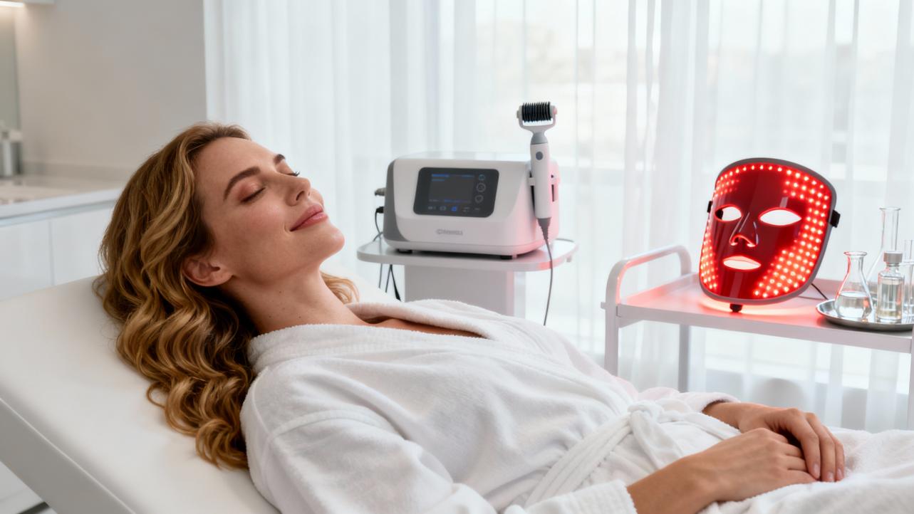
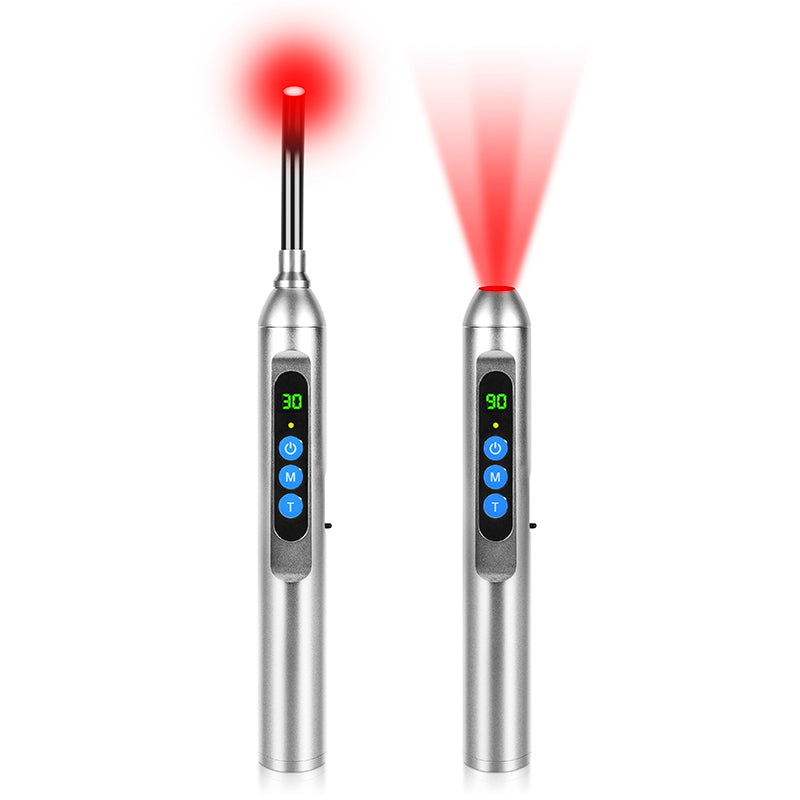
Red light therapy, often called low‑level light therapy or photobiomodulation, uses low‑energy red and sometimes near‑infrared wavelengths to change cellular behavior without burning or injuring tissue. Dermatology briefs from academic centers and the Cleveland Clinic describe it as non‑ionizing, non‑UV light, typically in the neighborhood of about 600 to 700 nanometers for red and about 800 to 900 nanometers for near‑infrared.
Mechanistically, the broader photobiomodulation literature, as summarized in PubMed‑indexed reviews, converges on the mitochondria. Chromophores such as cytochrome c oxidase absorb these wavelengths, mitochondrial respiration ramps up, adenosine triphosphate (ATP) production increases, and a controlled burst of reactive oxygen species and nitric oxide triggers downstream signaling. That signaling can upregulate collagen and elastin production, modulate inflammation, and nudge wound healing forward.
In skin‑specific trials and clinical experience, red light has repeatedly been shown to:
- Stimulate fibroblasts to produce more type I and III collagen and elastin, improving firmness and wrinkle depth.
- Reduce markers of inflammation and redness in conditions such as acne and rosacea.
- Support wound and scar remodeling by improving tissue repair and circulation.
- Improve overall tone and texture, including homogeneity of complexion, in anti‑aging studies.
One well‑characterized study published in a dermatologic journal and archived on PubMed Central tested a Dior x Lucibel red LED mask emitting around 630 nanometers, delivering approximately 15.6 joules per square centimeter in a 12‑minute session. Twenty middle‑aged volunteers used the mask twice a week for three months. Results showed progressive decreases in wrinkle depth and facial sagging, a nearly fifty percent increase in dermal density, and a substantial improvement in complexion homogeneity along with a reduction in oiliness and pore size. Importantly, these gains persisted for at least a month after stopping treatment, which supports true structural change rather than a transient plumping effect.
This is the biology and clinical backdrop: red light is not magic, but it clearly nudges skin cells into a more youthful, less inflamed state when dosed correctly. The question is how that intersects with post‑sun pigmentation.

Not All Light Colors Behave the Same
To understand red light’s role in pigment prevention, you have to zoom out to the full visible spectrum. The review on visible light and melanocyte biology divides visible light roughly into blue, green, yellow, and red bands, each with distinct targets and effects. Brand and clinic articles on LED therapy echo these differences when they talk about blue, green, red, and near‑infrared modes for different concerns.
Here is a simplified snapshot drawn from those sources.
Light band |
Rough range (nm) |
Main skin targets and effects related to pigment |
Blue visible |
About 400–490 |
Shallow penetration; activates OPN3 in melanocytes at higher doses; can trigger potent and long‑lasting hyperpigmentation, especially in darker phototypes; also used to kill acne bacteria. |
Green visible |
About 490–570 |
Primarily upper epidermis; in LED protocols from pigmentation‑focused brands, used to down‑regulate melanocytes and inhibit melanin production; reported to fade superficial dark spots and calm redness. |
Yellow visible |
About 570–595 |
Deeper effect on fibroblasts; in vitro work shows increased collagen I and antioxidant enzymes, and reduced UVA‑induced matrix‑degrading enzymes; framed as anti‑photoaging. |
Red visible |
About 630–700 |
Reaches the dermis; well‑documented for fibroblast activation, collagen and elastin support, antioxidant gene upregulation, reduced reactive oxygen species, and reversal of collagen degradation; used clinically for wrinkles, scars, and inflammatory skin conditions, and increasingly as an adjunct in pigmentation protocols. |
Near‑infrared |
About 800–900 |
Penetrates even deeper to subcutaneous tissues; some experimental work shows reduced tyrosinase and melanin, but other studies find stimulation of melanocytes in vitiligo; practiced estheticians highlight this mixed picture and often use near‑infrared cautiously in pigment‑driven disorders. |
In other words, blue and some green wavelengths can push melanocytes toward more pigment, while low‑dose yellow and red appear to protect dermal structure and oxidative balance. That distinction is crucial if you are choosing a post‑sun routine. For pigment‑prone skin, you do not want extra blue light blasts after an already intense day in the sun. You want wavelengths that resolve inflammation, support repair, and avoid overstimulating melanocytes.
How Red Light Might Prevent Post‑Sun Pigmentation
Dermatology researchers and estheticians do not yet have a large, controlled trial that looks at this exact scenario: group A spends a lot of time in the sun and then uses red light on a schedule, group B spends the same time in the sun but does not, and investigators track who develops more dark spots. That study has not been done. What we have instead are mechanistic studies, anti‑aging and hyperpigmentation trials, and clinical practice reports that together make a plausible case for red light as a post‑sun pigment‑control tool.
Calming the Post‑Sun Inflammatory Cascade
After a heavy sun day, your skin is not only tanned; it is inflamed and oxidatively stressed. Reactive oxygen species rise, inflammatory cytokines increase, and both keratinocytes and fibroblasts send distress signals. This environment is exactly what pushes melanocytes into overdrive, particularly around pre‑existing damage such as acne lesions, eczema patches, or subtle barrier compromise.
Photobiomodulation research, including the PubMed Central articles on red light and skin aging, shows that appropriately dosed red light can reverse part of that storm. By increasing ATP and modulating redox status, red light upregulates antioxidant pathways, decreases excessive reactive oxygen species, and reduces matrix‑degrading enzymes in fibroblasts. Clinical dermatology pieces from practices in Arizona and Pennsylvania highlight red light’s anti‑inflammatory effects in acne, rosacea, and post‑procedure care, reporting calmer skin, less redness, and faster resolution of lesions.
Mechanistically, this is the first route by which red light could reduce post‑sun pigmentation. If you can quiet inflammation and oxidative stress during the days after ultraviolet and blue light exposure, you remove some of the key triggers that tell melanocytes to dump more pigment into the upper skin layers. The data supporting this are indirect, but the biology is coherent.
Supporting Melanocytes Rather Than Shocking Them
Several hyperpigmentation‑focused LED brands, as well as an esthetician with more than fifteen years of experience featured by a red‑light device company, describe another effect: red light seems to normalize overactive pigment cells. Articles from companies specializing in red‑light face masks and panels state that longer red wavelengths can reach the basal layer where melanocytes live, and that repeated red‑light exposure may help “down‑regulate” overactive cells and balance melanin distribution.
An Australian device manufacturer frames this as red light “gently rebalancing” melanin production in melanocytes, both fading existing dark spots and lowering the odds of new ones when treatments are continued for several months. Another brand describes red light as strengthening the skin’s defense against ultraviolet damage, essentially pre‑conditioning the skin so it is less reactive the next time you go into the sun.
These reports are not randomized controlled trials, and a dermatology‑based hyperpigmentation expert emphasizes that red light should not be used as a stand‑alone cure. However, in real‑world protocols that combine pigment‑suppressing topicals, controlled exfoliation, and red light, both the clinician and her clients have seen meaningful normalization of blotchy pigment over time. Red wavelengths in the mid‑600s are her preference for pigment work, because the clinical literature on near‑infrared around 830 to 850 nanometers remains conflicted, with some experiments showing reduced melanin and others showing melanocyte stimulation.
From a prevention standpoint, the logic is this: if red light helps keep melanocytes in a calmer, more regulated state, then each episode of sun exposure is less likely to tip them into a full‑blown flare.
Making Texture and Tone More Uniform
Post‑sun pigment is not just a color problem; it is a structure problem. When dermal collagen is depleted and the skin surface is rough, any extra pigment catches the light and looks more obvious.
The Dior × Lucibel mask study is instructive here. Volunteers did not enroll for pigment specifically; they came in with general signs of aging. After three months of twice‑weekly red‑light sessions at a defined dose, investigators documented improvements in almost every structural parameter they measured. Crow’s‑feet wrinkles were reduced by more than a third. Sagging of the lower face decreased nearly a quarter. Dermal density rose close to fifty percent. Skin roughness and pore diameter fell substantially. The homogeneity of complexion, quantified by precise colorimetry between inner and outer cheek areas, improved by about a third. Sebum output dropped by more than two‑thirds, and pores laden with porphyrins, a proxy for congested, acne‑prone skin, fell as well. Every participant reported that their skin simply looked better.
Even though this trial did not separate “sun spots” from other color issues, a smoother, denser, better‑hydrated surface with more even light reflectance makes any residual pigment far less noticeable. In my own practice, I see this effect constantly: once the dermis is rebuilt and the surface is refined, clients often perceive their spots as “mostly gone” even when a close photograph shows that some pigment remains.

What the Evidence Really Supports
As a long‑time light‑therapy geek, I get excited by before‑and‑after images. But it is worth pausing for a sober evidence check. Academic summaries from the Cleveland Clinic, Stanford Medicine, and PubMed‑indexed photobiomodulation reviews all emphasize the same two points. First, red light clearly does something biologically real in skin. Second, the size and quality of clinical trials outside a few core indications remain modest.
For wrinkles, small randomized or controlled trials with red and near‑infrared panels have documented statistically significant but not miraculous improvements in roughness and elasticity over two to three months of regular use. For wound healing and scars, results are mixed, with some trials showing faster epithelialization and better cosmetic outcomes and others finding only short‑term differences that fade by around six weeks.
For hyperpigmentation specifically, the evidence base is still emerging. Several commercial and practice‑based articles describe positive outcomes in melasma and post‑inflammatory hyperpigmentation when red light is layered into broader protocols. An Australian device company reports that most users see visible fading of dark spots over four to six weeks of three to five sessions per week, with more marked changes after three months. A wellness brand positions red light as a gentle, non‑toxic option for acne‑related darkening and scars, supported by improved blood flow and reduced redness. An experienced esthetician calls red light a “must” element in pigment correction because of its healing and collagen‑supporting effects.
However, major medical centers remind readers that studies are often small, lacking placebo controls or standardized dosimetry, and long‑term safety data for consumer devices are still limited. Plus, there is no strong evidence that red light alone can prevent pigment in the absence of rigorous ultraviolet protection and pigment‑suppressing skincare. It is more accurate to consider red light a high‑leverage adjunct.
Designing a Pigment‑Smart Routine With Red Light
Even with those caveats, the convergence of mechanistic data, anti‑aging trials, and hyperpigmentation protocols suggests that red light deserves a place in a pigment‑conscious routine, especially after sun exposure. The key is to build it on top of strong fundamentals rather than in place of them.
First: Nail the Non‑Negotiables
Every dermatologist‑backed article in your research stack says the same thing in one way or another. If you want less pigment, you have to respect the sun. That means daily broad‑spectrum sunscreen of at least SPF 30 whenever you will be outside, avoiding deliberate tanning, and being realistic about midday summer exposure. Several of the hyperpigmentation‑focused articles also highlight the importance of not picking at spots or scabs, since mechanical trauma can turn a transient blemish into a lasting mark.
On the topical side, the esthetician featured by a medical‑grade red‑light brand lays out three pillars for treating hyperpigmentation: suppress melanin production, resurface or “re‑injure” the area in a controlled way so pigment can be lifted out, and use red light therapy to support healing and collagen. She singles out vitamin C at around twenty percent as a must‑have pigment inhibitor because it blocks tyrosinase, the key enzyme in melanin synthesis, and lightens dark spots while merely brightening normal skin. Other dermatologist‑authored pieces in your notes point to retinoids and antioxidants as additional tools for speeding cell turnover and limiting oxidative damage.
From a wellness‑optimization perspective, the lifestyle modifiers are not glamorous but they matter. Heavy alcohol intake is linked with more hyperpigmentation, likely through inflammation, liver burden, and increased ultraviolet sensitivity. Antioxidant‑rich foods, hydration, adequate sleep, and stress management all help the skin stay resilient, which means less over‑reaction when you do get incidental sun.
Then: Layer Red Light Thoughtfully After Sun
Now bring in the LEDs. The majority of clinical and practice‑based recommendations converge on a similar pattern: clean, dry skin; no sunscreen or occlusive products during the session; and regular treatments rather than sporadic blasts. Most dermatology clinics and device makers in your research suggest about ten to twenty minutes per session, performed several times per week, as the sweet spot. The Dior × Lucibel anti‑aging trial used twelve minutes twice weekly, spaced seventy‑two hours apart, and still delivered substantial structural gains.
For a pigment‑prone person coming off a day in intense sun, here is how I typically design a red‑light block in a routine while staying aligned with the published data. In the evening, after sun exposure, I have clients cleanse gently with a non‑stripping cleanser and skip exfoliating acids or retinoids if the skin feels at all tight or tender. Once the skin is clean and fully dry, they sit or lie in front of a red‑light device that uses predominantly mid‑600‑nanometer wavelengths, or they don a red‑light mask, for around ten to fifteen minutes. If their device manual recommends a specific distance, usually somewhere around a foot or less, they respect that rather than pressing the LEDs directly into the skin unless the product is designed for contact use.
The goal on these post‑sun evenings is not to chase a tan or force a “glow,” but to give the skin a calm, energy‑rich, low‑inflammation environment in which to repair. After the session, we layer pigment‑smart skincare: vitamin C in the morning, pigment inhibitors and, if the skin is not sunburned, appropriate retinoids at night, along with barrier‑supportive moisturizers. Sunscreen resumes the next day, and we continue red‑light sessions three to five times weekly for at least a month to ride out the delayed pigment phase.
The exact timing relative to sun exposure has not been rigorously studied, so I frame this as a strategy that is biologically plausible and grounded in existing red‑light dosing research, not as a proven medical protocol. For severe sunburns with blistering or systemic symptoms, the priority is medical care; at‑home light gadgets are not the first line in that situation.
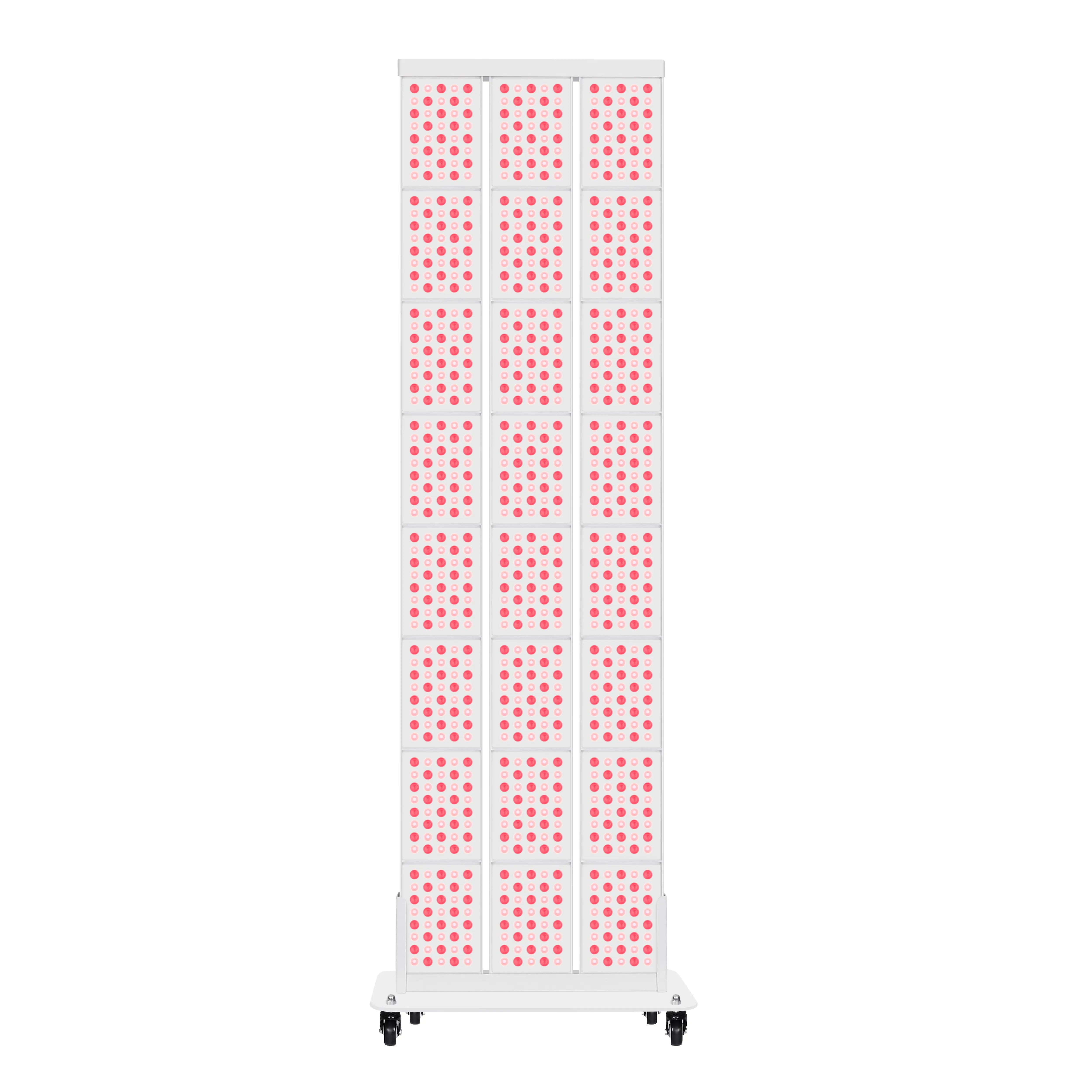
Respect Dose and Device Differences
One of the most important details in the Dior × Lucibel study is not the brand name; it is the dose. The authors point out the Arndt–Schulz law: in biological systems, too little stimulus produces no effect, an optimal middle range stimulates, and too much becomes inhibitory or even damaging. That is why they spaced sessions by about three days and why they optimized power density across the face rather than simply cranking up the total fluence.
Consumer devices vary enormously in wavelength accuracy, irradiance, and treatment time. Academic and clinical sources emphasize that at‑home devices are typically less powerful than clinic panels or laser‑based systems, which means they can be safer but also slower to produce change. This is one reason some people feel underwhelmed when they casually wave a low‑power wand around once a week. Consistency at an appropriate dose matters more than intensity.
For pigment‑prone users, there is another nuance. The esthetician quoted in the hyperpigmentation FAQ recommends sticking to red wavelengths in the mid‑600‑nanometer range for pigment work and being cautious with near‑infrared around 850 nanometers. Some studies at those longer wavelengths did reduce melanin production, but others used similar bands to stimulate melanocytes in vitiligo. Her practical advice is simple: if you have hyperpigmentation or melasma and your device allows you to turn off near‑infrared, do so and rely on red alone, at least initially.
Safety for Pigment‑Prone and Darker Skin
Overall, the safety profile of red light therapy is favorable when devices are used as directed. Academic overviews from the Cleveland Clinic and UCLA Health stress that red and near‑infrared light do not carry the DNA‑damaging risks of ultraviolet radiation. Side effects in trials and practice reports are usually limited to transient redness, tightness, or dryness. Serious complications are rare.
That said, several important cautions emerge in the research notes.
People with darker skin tones may be more sensitive to visible light‑induced pigmentation. The visible‑light review documents that blue and green wavelengths can generate stronger and more persistent pigment darkening in phototypes IV to VI than in lighter types. An article aimed at consumers from a medical center explicitly advises individuals with darker skin to be especially cautious with at‑home red‑light devices and to talk to a dermatologist first, because their skin may react differently and hyperpigmentation could theoretically worsen in some scenarios.
Blue light acne masks are a particular concern. Lifestyle‑oriented LED brands note that while most people do not see increased hyperpigmentation from their red and near‑infrared devices, individuals with freckles or strong pigment tendencies sometimes experience temporary darkening with pure blue light. One brand suggests that if blue light is used for acne in such users, it should be at low strength and followed immediately by red and near‑infrared sessions to help counteract pigment stimulation. For those whose main issue is pigmentation rather than acne, I generally recommend sidelining stand‑alone blue‑light devices and focusing on red.
Photosensitivity is another red flag. Several medical and brand articles remind readers that some medications, health conditions, and even topical ingredients can make the skin unusually reactive to light. Examples mentioned include certain antidepressants, nonsteroidal anti‑inflammatory drugs, antimalarials, and herbal supplements such as St. John’s wort. If you are in that category, any light‑based therapy, even non‑UV, should be cleared with your physician.
Finally, though red light devices intended for facial use are designed to minimize eye exposure, both academic and consumer safety guides recommend respecting device instructions for eye protection and never staring directly into bright LED panels.

Pros and Cons of Red Light for Post‑Sun Pigment Control
If you boil down all of this evidence and experience, red light therapy emerges as neither a miracle cure nor a gimmick. It is a useful lever with a distinct profile of strengths and limitations.
Aspect |
Potential advantages for pigment‑prone, sun‑exposed skin |
Limitations and cautions |
Mechanism |
Calms inflammation and oxidative stress after ultraviolet exposure; supports collagen and barrier repair, and may help normalize overactive melanocytes. |
Does not physically block ultraviolet or high‑energy visible light; cannot prevent damage from unprotected sun exposure. |
Evidence base |
Multiple clinical studies show improved skin quality, wrinkle reduction, and more uniform tone; practice reports and small studies suggest benefits in hyperpigmentation. |
Few large, well‑controlled trials specifically on pigment prevention; many hyperpigmentation claims are based on small or proprietary studies. |
Practical use after sun |
Non‑invasive, painless, and compatible with post‑sun skin when not severely burned; can be layered with sunscreen and pigment‑suppressing skincare. |
Optimal timing and dosing for post‑sun pigment prevention are not formally defined; overuse or underuse may blunt benefits. |
Device accessibility |
Wide range of at‑home masks, panels, and wands; FDA‑cleared options exist for skin and hair indications. |
Device power, wavelength accuracy, and quality vary widely; cheaper gadgets may underperform, while stronger units require more knowledge and care. |
Pigment‑type considerations |
Red‑only mid‑600‑nanometer devices have a strong safety record across skin types in anti‑aging trials; some studies intentionally included darker phototypes. |
Blue light and possibly some near‑infrared settings can aggravate pigmentation in certain conditions; darker skin, melasma, or vitiligo warrant individualized protocols and professional guidance. |
My Take as a Light‑Therapy Geek
If your main fear after a day in the sun is waking up a month later with new spots, red light therapy is one of the few at‑home interventions that can plausibly tilt the odds in your favor without adding toxic burden or downtime. The mitochondrial and anti‑inflammatory effects are real, the structural anti‑aging data are solid enough to matter, and clinical experience in hyperpigmentation is encouraging when red light is deployed as part of a broader, pigment‑smart strategy.
At the same time, the science is not nearly strong enough to justify skipping sunscreen, ignoring shade, or throwing away your vitamin C and retinoids. The smartest move is to think of red light as a way of helping your skin process the sun more gracefully, not as a license to roast. In my own protocols, the people who win long term are the ones who treat red light like a daily or near‑daily self‑care habit layered onto boring fundamentals, not as an emergency fix after every sun binge.
Used that way, with realistic expectations and a respect for dose, red light can absolutely be one of your highest‑leverage tools for keeping post‑sun pigmentation from writing a permanent story on your skin.
Frequently Asked Questions
Does red light therapy replace sunscreen for preventing dark spots?
No. Red light therapy does not block ultraviolet or blue light and cannot prevent the initial DNA damage and oxidative stress that trigger pigment. Academic and clinical sources are very clear that daily broad‑spectrum sunscreen, sun‑smart behavior, and evidence‑based topicals are the primary tools for preventing hyperpigmentation. Red light is best used as a supportive therapy to help your skin recover and remodel more cleanly after inevitable sun exposure.
How quickly can red light change post‑sun pigmentation?
Brand reports and clinical observations suggest a pattern of subtle improvement within about one to two weeks of consistent use and more visible fading of deeper discoloration after four to twelve weeks. The Dior × Lucibel trial measured meaningful changes in wrinkles, firmness, and complexion homogeneity after one month, with further gains at two and three months. For hyperpigmentation, most device makers and practitioners recommend treating at least three months before judging full results, and maintaining a regular schedule thereafter.
Is near‑infrared safe if I have melasma or very dark skin?
The answer is nuanced. Some laboratory studies using near‑infrared wavelengths around 830 to 850 nanometers show decreased tyrosinase activity and melanin production, which sounds good for hyperpigmentation. Other studies, however, use similar wavelengths to stimulate melanocytes in vitiligo, essentially doing the opposite. Because of this mixed picture, an experienced pigmentation‑focused esthetician featured by a red‑light company chooses to rely primarily on mid‑600‑nanometer red light for hyperpigmentation work and suggests turning off or minimizing near‑infrared in clients who are pigment‑prone. If you have melasma or very dark skin and your device includes near‑infrared, it is worth discussing settings and strategy with a dermatologist or knowledgeable practitioner rather than guessing.
Can red light therapy itself cause hyperpigmentation?
For pure red‑light devices that do not emit ultraviolet or high doses of shorter visible wavelengths, the available data and clinical experience suggest that hyperpigmentation is unlikely in most users when devices are used as directed. Some medical sources even note that red and near‑infrared LEDs can decrease the appearance of pigment and improve uneven tone. That said, there are important exceptions. Blue‑heavy devices can darken skin in some individuals, especially with repeated exposure and in darker phototypes. People on photosensitizing medications or with particular skin diseases may also react unpredictably. This is why dermatology organizations and hospital‑based experts consistently recommend checking with a dermatologist before starting red‑light therapy if you have significant pigment issues, darker skin, or complex medical history.
References
- https://pmc.ncbi.nlm.nih.gov/articles/PMC10311288/
- https://med.stanford.edu/news/insights/2025/02/red-light-therapy-skin-hair-medical-clinics.html
- https://my.clevelandclinic.org/health/articles/22114-red-light-therapy
- https://www.gundersenhealth.org/health-wellness/aging-well/exploring-the-benefits-of-red-light-therapy
- https://www.uclahealth.org/news/article/5-health-benefits-red-light-therapy
- https://www.aad.org/public/cosmetic/safety/red-light-therapy
- https://franklinderm.net/anti-aging/red-light-therapy-for-skin-health-and-anti-aging-what-the-research-shows/
- https://lansdaleplasticsurgery.com/red-light-therapy-benefit
- https://www.englishdermatology.com/the-power-of-red-light-therapy-for-healthier-skin/
- https://higherdose.com/blogs/the-high-life-blog/red-light-therapy-for-hyperpigmentation-how-does-it-work?srsltid=AfmBOooA_CZxOPw49WFbg5AvyJrGDMQ40nQ_bmjO0ge23zuyIXLMGpyu