
Why Hypertrophic Scars Are So Hard To Soften
If you have a raised, tight, stubborn scar that refuses to flatten no matter how much you massage, moisturize, or tape it, you are dealing with hypertrophic scarring. These scars form when your body repairs a deep wound by laying down collagen-rich fibrous tissue that is thicker and tougher than the surrounding skin. Unlike keloids, hypertrophic scars usually stay within the borders of the original wound, but they can still be raised, firm, red, itchy, and sometimes restrictive if they cross joints or sit on high‑tension areas.
Research summarized by academic groups such as the University of California describes hypertrophic and keloid scars as part of skin fibrosis: abnormal wound healing with excess fibroblast proliferation and collagen deposition. One review notes that fibrotic scarring affects more than one hundred million people in developed countries each year and fuels a scar-treatment market worth around twelve billion dollars annually in the United States alone. That is a lot of people quietly fighting thick, uncooperative scars.
In practice, hypertrophic scars are a “Goldilocks failure” of healing. You want just enough fibroblast activity and collagen to close the wound and restore strength, but not so much that the repair overshoots and hardens into ropey tissue. Traditional treatments like silicone sheeting, pressure garments, steroid injections, and pulsed dye laser all try to nudge the biology back toward that sweet spot. Over the last decade, red and near‑infrared light have joined that conversation as a gentle way to modulate fibroblasts, inflammation, and blood flow without burning or cutting the skin.
As someone who has spent years experimenting with photobiomodulation devices on my own surgical and acne scars, and who reads the underlying papers rather than just the marketing, I view red light therapy as a promising adjunct for hypertrophic scar softening. The key word is adjunct. It is not a magic eraser, but it can be a surprisingly effective supporting actor when you respect the physics, the biology, and the limits of the current evidence.
How Red and Near‑Infrared Light Interact With Scar Tissue
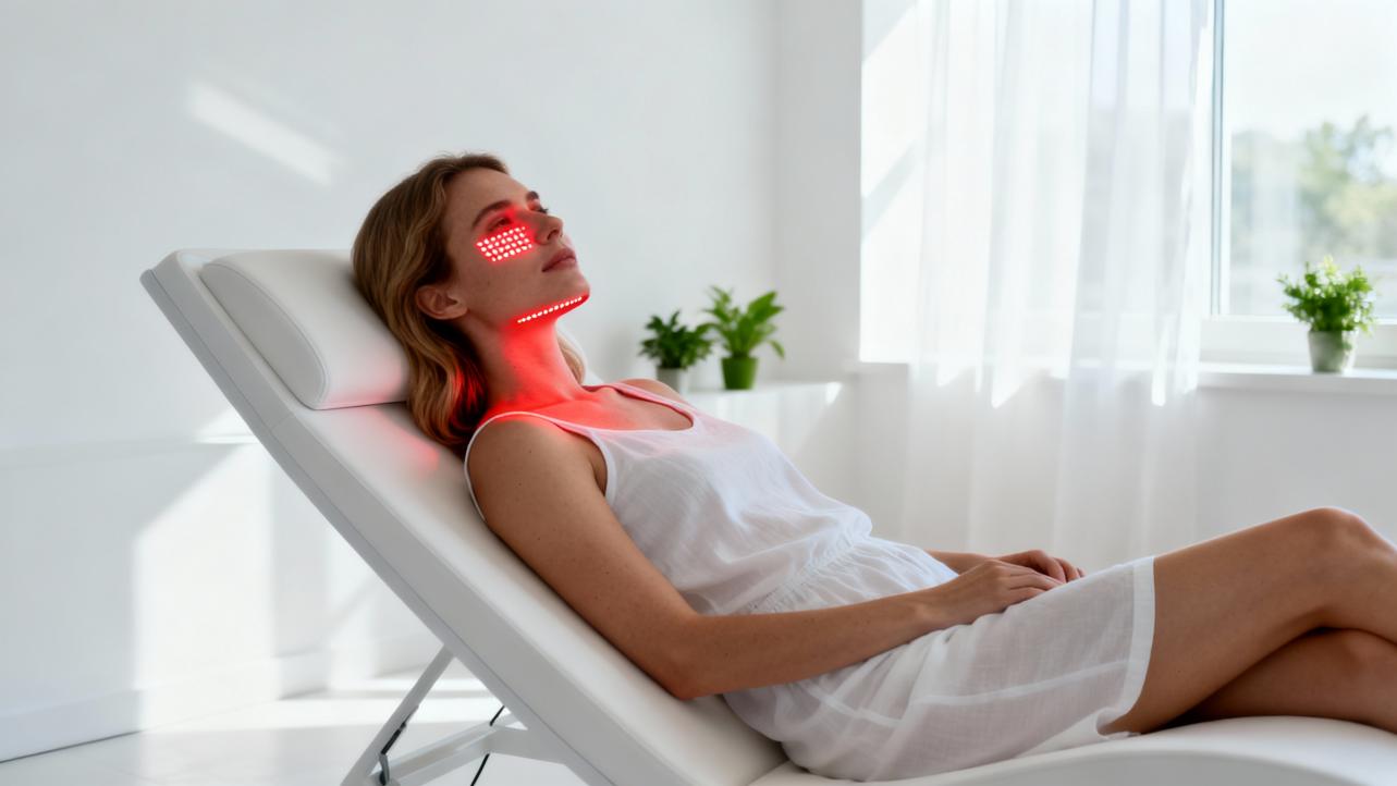
Cleveland Clinic describes red light therapy as the use of low‑level red or near‑infrared light to act on mitochondria, the “power plants” of cells. By nudging mitochondrial enzymes, red light can increase cellular energy production, stimulate fibroblasts, promote collagen synthesis, enhance local blood circulation, and reduce inflammation. Importantly, this is non‑thermal. The doses used in photobiomodulation do not aim to heat or destroy tissue the way ablative lasers do.
A randomized controlled trial in cosmetic dermatology, published in a peer‑reviewed journal and summarized in the research notes above, illustrates this nicely. In that study, one hundred thirty‑six volunteers received red and near‑infrared light treatments with doses around eight to ten joules per square centimeter in the red band per session. Over thirty sessions, they experienced measurable increases in intradermal collagen density and smoother skin texture, with no clinically meaningful ultraviolet exposure and no serious adverse events. That trial focused on wrinkles rather than scars, but it proves a core point: appropriately dosed red light can remodel dermal collagen in living humans without injury.
Stanford Medicine experts frame modern red light applications under the umbrella of photobiomodulation, a term formally introduced into biomedical indexing in 2015 and now supported by hundreds of studies. Mechanistically, they highlight that light at specific wavelengths can trigger vasodilation, alter inflammatory signaling, and influence hair follicles or dermal structure. For wound healing and scars specifically, they note that the data are mixed. Some trials show faster healing early on with red light and even healing times cut roughly in half in certain models, but those differences sometimes fade out by six weeks, and not every study finds statistically significant cosmetic gains.
From a scar perspective, that nuance matters. If a modality primarily accelerates early closure and calms inflammation, it may change how much fibrotic tissue your body lays down and how rigid it becomes. At the same time, if long‑term differences are modest or inconsistent, you should expect subtle softening and better blend with surrounding skin rather than dramatic erasure.

Fibroblasts, Collagen, and the “Just Enough” Problem
At the heart of hypertrophic scar softening is the fibroblast. These cells are your collagen factories. In normal healing, they ramp up, deposit support structures, then taper off as the wound matures. In hypertrophic and keloid scarring, fibroblasts stay too active and keep producing and organizing collagen in a way that raises and stiffens the scar.
An in vitro study summarized in the notes explored what happens when cultured dermal fibroblasts are exposed to low‑level infrared light from light‑emitting diodes. Under certain wavelength and dose combinations, fibroblast proliferation decreased compared with non‑irradiated controls. The cells did not show signs of gross injury or widespread death; the light was not burning them. Instead, it appeared to be dialing down their growth. The authors emphasized that this effect was dose‑dependent, which fits a wider theme in photobiomodulation: some doses stimulate cell activity, while others inhibit it.
Researchers at the University of California went further, translating this concept into a patented strategy for treating keloids and other fibrotic skin conditions. Their data indicate that carefully chosen red and infrared LED doses can reduce fibroblast proliferation and migration, two behaviors central to raised, invasive scars. The approach is explicitly described as non‑invasive, non‑ultraviolet, and ultimately intended for safe home use with affordable, portable devices.
Taken together, the petri dish and patent work suggest a compelling model. Early or established hypertrophic scars are essentially fibroblast‑driven “overhealing.” If you can use red or near‑infrared light to gently tell those fibroblasts to calm down—without damaging the surrounding tissue—you may end up with a softer, flatter, more pliable scar over time. That is the biological promise behind using red light to soften hypertrophic scars.
Clinical Evidence: The CURES Trial On Surgical Scars
The cleanest clinical test we have so far for red light’s impact on early hypertrophic‑prone scars is the CURES trial, a phase II study conducted at an academic center and registered as NCT03795116. In this randomized, temperature‑matched, single‑blind, split‑face trial, thirty adult facelift patients each served as their own control. One side of their periauricular incision received high‑fluence red LED therapy, while the opposite side received a mock treatment.
Treatments began roughly one week after surgery, between postoperative days seven and ten, once early healing was underway but scarring was still very immature. Patients were divided into three groups of ten, each assigned a different fluence: one hundred sixty, three hundred twenty, or four hundred eighty joules per square centimeter. All groups used the same wavelength, a narrowband red LED centered around six hundred thirty‑three nanometers, delivered with a device positioned within about ten millimeters of the skin.
Each participant received three in‑office sessions per week for three weeks, for a total of nine treatments. The primary endpoint was objective scar pliability, measured as induration using a SkinFibroMeter. Investigators also collected Patient and Observer Scar Assessment Scale scores, which rate aspects like thickness, color, stiffness, and overall impression on a scale where one represents normal skin and ten represents the worst imaginable scar. Additional instruments assessed dermal collagen and water content.
The headline result is sobering but important. Across the full cohort, there was no statistically significant difference in quantitative scar pliability between LED‑treated and mock‑treated sites at the primary time point. In other words, in this well‑designed split‑face study, red light did not clearly win on its main endpoint. However, at certain fluence levels, there were signals that treated scars improved more from baseline than controls in observer‑rated scales and in induration. Those trends suggest a possible dose‑dependent benefit, but they were not strong enough to definitively claim red light as a proven anti‑scarring therapy.
On safety, the news is better. Across this trial and earlier phase I studies on normal skin (often called STARS 1 and 2, with more than one hundred participants combined), doses up to four hundred eighty joules per square centimeter at six hundred thirty‑three nanometers were generally well tolerated. In the facelift population, treatment‑site adverse events were limited to mild blistering in two patients and mild swelling in one, all resolving without long‑term problems. Crucially, participants also underwent a pre‑enrollment photosensitivity test, and individuals with photosensitizing medications, light‑sensitive diseases, diabetes, lupus, coagulation disorders, tobacco use, or local skin disease were excluded.
The practical takeaway is that high‑fluence red LED therapy appears safe in the early post‑surgical period for appropriately screened patients and may modestly improve some aspects of scar pliability and appearance at the right dose. At the same time, the lack of a clear, statistically robust primary effect means we cannot yet call this an established standard of care for hypertrophic scar prevention or softening. It is a promising adjunct, not a replacement for conventional scar management strategies.

Wound Healing And Scar Biology Under Light
If you zoom out from surgical facelifts to wound healing more broadly, a pattern emerges. Several lines of evidence now show that red and near‑infrared light can meaningfully influence how skin repairs itself, which is directly relevant to how a hypertrophic scar forms and evolves.
A University at Buffalo–led study examined radiation‑induced skin damage, a condition called radionecrosis that behaves a lot like a deep burn with inflammation, impaired blood flow, and scarring. In an animal model, researchers used low‑dose red and near‑infrared LED photobiomodulation as an adjunct to standard wound care. Without light therapy, radionecrosis wounds took about sixty‑one days to close. Near‑infrared treatment shortened this to around forty‑nine days, and red light therapy brought closure down to roughly forty‑two days. That amounts to wound healing up to about fifty percent faster, alongside reductions in inflammation and improved circulation. The same group’s earlier work showed that photobiomodulation can activate TGF‑beta 1, a protein that stimulates fibroblasts and macrophages to coordinate repair.
Another recent paper in a Nature family journal, titled “Visible light accelerates skin wound healing and alleviates scar formation in mice by adjusting STAT3 signaling,” adds more mechanistic insight. The authors report that non‑ultraviolet visible light exposure in mouse models sped up cutaneous wound closure and reduced subsequent scar formation. They tie this effect to modulation of STAT3, a transcription factor that integrates inflammatory and growth factor signals and has known roles in skin remodeling and fibrosis. The reference list of that paper reads like a map of wound healing biology, highlighting macrophage phenotypes, monocyte chemoattractant protein‑1, and pathways that connect mechanical force and inflammatory signaling to skin fibrosis.
When you layer that on top of classic low‑level light therapy research, which shows that red and near‑infrared wavelengths in the six hundred to eight hundred nanometer range can stimulate or inhibit cell proliferation depending on dose, a coherent narrative emerges. Properly dosed red or near‑infrared light can accelerate the early phases of healing, modulate immune cell behavior, shape angiogenesis, and adjust fibroblast activity through pathways that include STAT3 and mitochondrial reactive oxygen species. In animal models, that can translate into both faster wound closure and reduced scarring.
The caution is that not every benefit observed in mice or rats automatically appears in human hypertrophic scars. Skin thickness, tension, immune context, and the sheer time scale of human scarring are different. But when I look across animal data, in vitro fibroblast studies, and human photobiomodulation trials, I see enough convergence to justify clinical curiosity. Red light therapy has real biological effects that plausibly intersect with the core drivers of hypertrophic scarring.
What Red Light Therapy Can Realistically Do For Hypertrophic Scars Today
Where does all of this leave a person staring down a raised surgical or traumatic scar and wondering if parking in front of a red light panel will help? The fairest answer, based on the evidence summarized by Cleveland Clinic, Stanford Medicine, Harvard Health, and multiple clinical and preclinical reports, is that red light therapy is a low‑risk, potentially helpful adjunct that may soften and blend hypertrophic‑prone scars, especially when used early and consistently, but it is not a stand‑alone cure.
Clinically, dentists and surgeons are already integrating red or infrared therapy around oral and facial scars. One dental practice reports using infrared light as a core element of post‑surgical scar care, with observed reductions in scar thickness, discoloration, and improvements in tissue elasticity and softness. Another clinic describes red light as a gentle adjunct after dental surgery or temporomandibular joint procedures to reduce pain and inflammation and help visible scars mature more favorably. Patients often report smoother skin and less conspicuous scars after a series of sessions that typically last ten to twenty minutes and continue weekly for several weeks.
Aesthetic practices and home‑use brands describe similar patterns in acne, injury, and cosmetic surgery scars. They highlight combined red and near‑infrared wavelengths, often around six hundred thirty‑three and eight hundred thirty nanometers, to stimulate fibroblasts, increase blood flow, and activate “wound healing cells” several centimeters below the surface. Users commonly notice subtle improvements in skin texture and scar color after four to six weeks of consistent use, with more visible softening taking eight to twelve weeks or longer.
However, independent assessment from Harvard dermatology experts points out that the overall evidence for consumer LED devices in scarring and photoaging is still limited. Many studies are small, short‑term, and lack robust control groups. Device quality and dosing parameters vary widely, and Food and Drug Administration clearance usually speaks to safety rather than proven efficacy. Cleveland Clinic echoes this, emphasizing that while red light is promising for wrinkles, scars, and inflammatory skin conditions, there is not yet enough high‑quality data to support many of the sweeping health claims being made online.
In practice, that means you should expect red light therapy to be incremental and synergistic. When hypertrophic scars soften, it is often because you are combining several levers: pressure or tension control, silicone or occlusive hydration, targeted lasers for redness, and perhaps steroid injections when appropriate. Red light therapy fits as a non‑invasive, low‑pain tool that can support deeper biology while you keep doing those established things.

How I Integrate Red Light Around Hypertrophic Scars
In my own use and in protocols I design for clients who are working with their physicians, I treat red light as a structured intervention, not a casual gadget. The research points to three variables you can control: timing relative to the wound, the light dose, and the overall scar‑care environment.
For timing, several sources converge on starting light treatment once the wound is closed, sutures are removed, and your surgeon clears it. In the facelift trial, red light began about a week after surgery. Dental and aesthetic clinics also describe starting early, within the first days to weeks after closure, to influence how the scar forms rather than waiting until it is fully mature. If you are dealing with an older hypertrophic scar, you can still use red light, but you should expect slower and perhaps smaller changes. Consumer research summaries make it clear that mature scars can respond, but they often need months of consistent treatment.
For dose and schedule, I like to align with ranges that show up across both clinical and consumer contexts. The facelift trial used high fluences at six hundred thirty‑three nanometers three times per week for three weeks. Skin rejuvenation studies used about eight to ten joules per square centimeter in the red band per session, delivered over twelve to twenty‑five minutes, twice weekly for fifteen to thirty sessions. Home device guidance from manufacturers who base their recommendations on the literature typically suggests sessions of ten to twenty minutes, three to five days per week, with the device around six to twelve inches from the skin. Users often start to see early textural shifts after a month, with more meaningful changes requiring two to three months.
The table below synthesizes the dosing patterns described in the research notes and in clinical practice reports. These are not rigid prescriptions, but they illustrate how real‑world use clusters around certain wavelengths and schedules.
Setting or Study Context |
Wavelengths and Light Type |
Typical Session Parameters |
Frequency and Duration |
Scar‑Relevant Notes |
Facelift CURES trial (post‑surgical facial scars) |
Narrowband red LED around 633 nm |
Fluence 160, 320, or 480 J/cm² per session |
Three times per week for three weeks, starting about 7–10 days after surgery |
Safe in screened patients; no significant effect on primary pliability endpoint; some dose‑dependent trends toward improved scar scores |
Dental and facial surgery clinics |
Red and near‑infrared LEDs; specific wavelengths often centered near 630–830 nm |
Sessions usually 10–20 minutes on the scar region |
Weekly or multiple times per week over several weeks |
Reports of smoother, less visible post‑surgical scars and reduced pain and inflammation; considered a gentle adjunct |
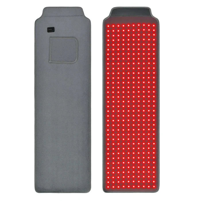
Home protocols for scars and skin texture |
Red and near‑infrared light between roughly 630–850 nm |
Sessions 10–20 minutes with device about 6–12 inches away |
About 3–5 sessions per week; subtle changes after 4–6 weeks, more visible changes after 8–12 weeks |
Users and providers describe gradual softening, improved texture, and better blending of acne, surgical, and stretch‑related scars |
The third pillar is the surrounding scar‑care environment. A detailed overview of scars and stretch marks notes that no topical or device completely removes scars. Evidence‑based topical options include silicone gels or sheets to hydrate and flatten raised scars, prescription retinoids to boost cell turnover in certain atrophic or early stretch marks, and corticosteroid creams or injections to reduce inflammation in raised scars. In‑office procedures such as vascular and fractional lasers, microneedling, chemical peels, and dermabrasion can all remodel scar tissue under professional guidance. Surgical revision and subcision are reserved for more severe, tethered, or function‑limiting scars.
When I build a plan, I stack red light alongside these tools rather than instead of them. For example, after your surgeon clears you, you might apply silicone sheeting during the day for tension and hydration, use gentle massage, attend pulsed dye laser sessions if redness is prominent, and integrate red light therapy three to five evenings per week. The red light sessions focus on modulation of fibroblasts and inflammation while the other treatments manage mechanical forces, vascular changes, and epidermal turnover.

Safety, Caution, And Who Should Think Twice
In terms of safety, the convergence of data is reassuring when red light is used correctly. Cleveland Clinic, Harvard Health, Stanford Medicine, and multiple clinical trials all describe red and near‑infrared light therapy as non‑invasive, non‑ultraviolet, and generally well tolerated. Short‑term use within recommended parameters is not associated with serious adverse effects in the published studies cited here. Animal work on radiation wounds even suggests that photobiomodulation can improve healing without stimulating tumor cells, likely because cancer cells have altered metabolic pathways.
There are still important caveats. Investigators in the facelift trial screened out participants with photosensitizing medications, light‑sensitive diseases, diabetes, lupus, coagulation problems, tobacco use, or local skin disease, and they performed a forearm photosensitivity test before enrolling. Consumer‑facing centers and academic hospitals alike advise people with medical conditions or those on photosensitizing drugs to consult their dermatologist or surgeon before starting red light therapy. The same applies if you have a history of skin cancer or a suspicious lesion near the treatment area.
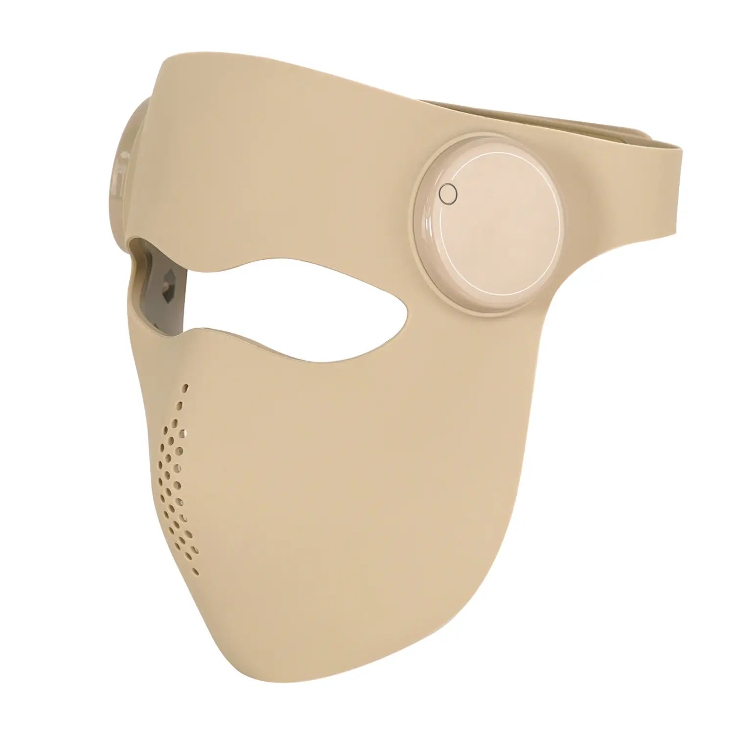
Eye protection is non‑negotiable. Harvard dermatologists highlight a real‑world recall of a consumer acne mask after concerns about potential eye damage in vulnerable populations. Even though home devices are generally lower power than clinic systems, you should shield your eyes and avoid staring directly into the LEDs.
Finally, the long‑term safety of chronic, high‑frequency cosmetic LED exposure is not fully known. Available data are encouraging, but most studies are months long, not decades. From a wellness‑optimizer standpoint, that argues for using enough light to get a plausible benefit, but not turning your bedroom into a permanent photobiomodulation chamber for hours every day without medical oversight.
FAQ: Hypertrophic Scars And Red Light Therapy
Can red light therapy completely flatten a hypertrophic scar?
No current therapy, including red light, reliably erases hypertrophic scars. Evidence and clinical experience suggest that red light can contribute to softer, more pliable, and less conspicuous scars over time, especially when combined with established treatments like silicone, pressure, and targeted lasers. The CURES trial and expert reviews show modest or mixed improvements rather than dramatic transformations. Think in terms of percentage improvements and better texture and comfort, not a return to perfectly unscarred skin.
Is red light therapy more useful for fresh scars or old ones?
The best mechanistic rationale and the most structured clinical data sit around early scars, in the weeks and first few months after surgery or injury, when fibroblasts and inflammatory pathways are still actively shaping the scar. That is when high‑fluence red light has been tested in facelift patients, and when dentists and surgeons tend to add red or infrared sessions. Older hypertrophic scars can still respond, but they usually need longer courses and the changes tend to be slower and subtler.
How long before I notice changes in a hypertrophic scar?
Consumer protocols drawn from the research note that many people start noticing subtle changes in overall skin texture and tone after around four to six weeks of regular red light use, with more visible softening and blending often appearing after eight to twelve weeks or longer. Given that scar remodeling is a slow biological process measured in months, not days, it is reasonable to commit to at least two to three months of consistent use before judging the full effect, ideally under the guidance of a dermatologist or surgeon who can also adjust other aspects of your scar care.
Could red light therapy make my scar worse or trigger cancer?
The available data do not show red or near‑infrared light worsening scars when used within recommended dosing ranges. Trials in wound healing and radionecrosis models actually show better quality healing and less severe scarring. An animal study from University at Buffalo suggests that photobiomodulation speeds healing without stimulating tumor growth, likely because tumor cells respond differently to these light‑induced pathways. That said, red light therapy has not been exhaustively tested in every scenario, and people with a history of skin cancer, pre‑cancerous lesions, or on certain medications should work closely with their physicians to decide what is appropriate.
Closing Thoughts From A Light Therapy Geek
Hypertrophic scars are a classic case where biology overshoots, and red light therapy gives us a nuanced way to nudge, not bludgeon, that biology back toward balance. The science so far tells a consistent story: red and near‑infrared light can accelerate healing, modulate fibroblasts, and sometimes yield softer, better‑behaving scars, but the effects are modest, dose‑dependent, and strongest when paired with smart, conventional care. If you treat red light therapy as a disciplined, evidence‑aware tool rather than a miracle cure, it can become a powerful ally in softening the scars your body insists on overbuilding.
References
- https://lms-dev.api.berkeley.edu/red-light-skin-treatment
- https://digitalcommons.cedarville.edu/cgi/viewcontent.cgi?article=1013&context=education_theses
- https://clinicaltrials.gov/study/NCT03795116
- https://www.health.harvard.edu/diseases-and-conditions/led-lights-are-they-a-cure-for-your-skin-woes
- https://techtransfer.universityofcalifornia.edu/NCD/23101.html
- https://pmc.ncbi.nlm.nih.gov/articles/PMC3926176/
- https://dev.ppc.uiowa.edu/libweb/5P8050/HomePages/RedLightTherapyForScarTissue.pdf
- https://www.med.umich.edu/1libr/PMR/Lymphedema/LymphTherapyTechniques.pdf
- https://www.bcm.edu/healthcare/specialties/dermatology/laser-surgery/laser-treatment-with-pulsed-dye-laser
- https://www.buffalo.edu/news/releases/2022/01/029.html